. 2025 Feb 21;15(1):6334.
doi: 10.1038/s41598-025-90805-7.

Adipose-derived stem cells using fibrin gel as a scaffold enhances post-hepatectomy liver regeneration

Affiliations

Adipose-derived stem cells using fibrin gel as a scaffold enhances post-hepatectomy liver regeneration

Hiroki Imamura et al. Sci Rep. .

Erratum in

Abstract

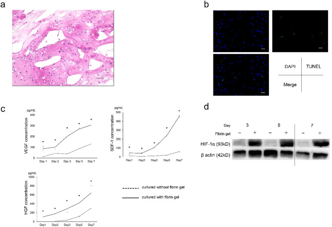
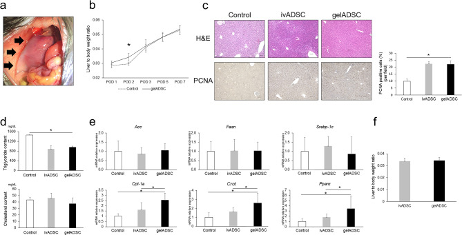
We investigated the potential of adipose-derived stem cells (ADSCs) in preventing post-hepatectomy liver failure, emphasizing the necessity of direct administration using a scaffold. A fibrin gel scaffold was employed for ADSCs (gelADSC) to assess their therapeutic impact on liver regeneration in both in vitro and in vivo settings. Experiments were conducted on C57BL/6 mice with normal livers and those with chronic hepatitis. We also explored the role of extracellular vesicles (EVs) secreted by ADSCs in conjunction with fibrin gel. GelADSC showed sustained release of hepatocyte growth factor, vascular endothelial growth factor, and stromal cell-derived factor 1 for at least 7 days in vitro. In vivo, gelADSC significantly enhanced postoperative liver regeneration by upregulating the cell cycle and fatty acid oxidation in both normal and chronically hepatitis-affected mice. The therapeutic effects of gelADSC were potentially favorable over those of intravenously administered ADSCs, especially in mice with chronic hepatitis. Increased EV secretion associated with fibrin gel use was significantly linked to enhanced liver regeneration post-surgery through the promotion of fatty acid oxidation. The findings underscore the enhanced therapeutic potential of gelADSC, particularly in the context of chronic hepatitis, possibly compared to intravenous administration.

Keywords: Adipose-derived stem cell; Fibrin gel; Hepatectomy; Liver regeneration; Paracrine effect.


Conflict of interest statement

Declarations. Competing interests: HI received a research fund from Rohto Pharmaceutical Co., Ltd., and SK (Shogo Kobayashi) received honoraria from AstraZeneca and Taiho. The other authors (YT, AH, SK (Shunbun Kita), KS, YI, DY, TN, HT, DH, TK, KT, TK, SS, IS, SM, YD, HE) declare no competing financial interests.

Figures

Fig. 1
Fig. 1
In vitro evaluation of ADSCs embedded in fibrin gel. (a) H&E staining of gelADSC after 7-day incubation exhibited numerous ADSCs remaining in the fibrin gel. Scale bar = 50 μm. (b) TUNEL staining of gelADSC after 7-day incubation revealed viable ADSCs. Scale bar = 50 μm. (c) ADSCs embedded in fibrin gel (solid line) secreted significantly higher amounts of VEGF, HGF, and SDF-1 than did ADSCs incubated without fibrin gel (dotted line). (d) Western blot analysis showing the enhanced expression of HIF-1α by ADSCs embedded in fibrin gel on days 3, 5, and 7. *p < 0.05.
Fig. 2
Fig. 2
In vivo evaluation of ADSCs embedded in fibrin gel. (a) H&E staining of gelADSC on postoperative day 7 revealed numerous ADSCs remaining in the fibrin gel. Scale bar = 50 μm. (b) TUNEL staining of gelADSC on postoperative day 7 revealed viable ADSCs. Scale bar = 50 μm. (c) Immunostaining of gelADSC using antibodies for CD90, CD105, arginase-1, and hepatocyte-specific antigen (HSA) revealed that ADSCs in the fibrin gel were diffusely positive for CD90 and CD105 but negative for arginase-1 and HSA. Scale bar = 50 μm. (d) Berlin staining of ADSCs confirmed labeling with iron. Scale bar = 50 μm. (e) The distribution of ADSCs in vivo was assessed by T2WI MRI. Fibrin gel exhibited a high-density signal on the liver surface (yellow arrows), whereas it exhibited a low-density signal when iron-labelled ADSCs were embedded in it (blue arrows). Intravenous administration (ivADSC) resulted in a hepatic distribution of ADSCs, as evidenced by the low-density liver parenchyma (red arrows).
Fig. 3
Fig. 3
Therapeutic effect of gelADSC after hepatectomy for normal liver. (a) Intraoperative photograph showing gelADSC placed on the surface of the remnant liver (black arrows). (b) The liver to body weight ratio was significantly higher on POD 2 in the gelADSC group than in the control group. (c) Representative images of the histological evaluation of remnant liver showing the accumulation of lipid in the control group on H&E staining, whereas such accumulation is decreased in the ivADSC and gelADSC groups. Immunohistochemistry for PCNA showed a significant increase in PCNA-positive cells in the gelADSC group compared with the control group. Scale bar = 100 μm. (d) A triglyceride assay revealed that the liver triglyceride content in the gelADSC group was significantly higher than in the control group but comparable to the ivADSC group. The cholesterol content was comparable between the three groups. (e) mRNA expression in the liver was evaluated by real-time PCR. The expression of Acc, Fasn, and Srebp-1c was comparable between the three groups, whereas that of Cpt-1a, Crot, and Pparα was significantly upregulated in the gelADSC group, the ivADSC group, and the control group in that order. (f) The liver to body weight ratio on POD 2 was comparable between the gelADSC group and the ivADSC group. *p < 0.05.
Fig. 4
Fig. 4
Therapeutic effect of gelADSC after hepatectomy for chronic hepatitis. (a) Masson trichrome staining of the resected liver confirmed liver fibrosis. Scale bar = 100 μm. (b) The liver to body weight ratio gradually increased in both the control and the gelADSC groups, with a significant difference on POD 3 in favor of the gelADSC group, although the difference was not significant at PODs 1, 2, 5, and 7. (c) Representative H&E images of remnant liver demonstrating lipid accumulation in the control group, with less lipid accumulation in the ivADSC and gelADSC groups. Immunohistochemistry for PCNA showed a significant increase of PCNA-positive cells in the gelADSC group compared with the control and ivADSC groups. Scale bar = 100 μm. (d) mRNA expression in the liver was evaluated by real-time PCR. The expression of Acc, Fasn, and Srebp-1c was comparable between the three groups, whereas that of Cpt-1a, Crot, and Pparα was significantly upregulated in the gelADSC group compared with the control group and the ivADSC group. (e) The liver to body weight ratio at POD 3 was significantly higher in the gelADSC group than in the ivADSC group. *p < 0.05.
Fig. 5
Fig. 5
MRI evaluation of ADSCs administered in a chronic hepatitis model. (a) The distribution of ADSCs administered in a chronic hepatitis model was evaluated by T2WI on PODs 1, 3, and 7. In the gelADSC group, fibrin gel exhibited low-intensity enhancement, reflecting the presence of ADSCs labeled with iron. In the ivADSC group, the distribution of intravenously administered ADSCs was not apparent. (b) The liver signal intensity relative to that of muscle was evaluated in the ivADSC groups with normal liver and with chronic hepatitis using the ImageJ software. The intensity was significantly lower in mice with hepatectomy and normal liver than in those with chronic hepatitis on POD 1, 3, and 7. *p < 0.05.
Fig. 6
Fig. 6
Evaluation of the impact of extracellular vesicles (EVs) secreted from ADSCs. (a) EV secretion increased in a time-dependent manner, both with and without fibrin gel. Furthermore, EV secretion was higher with the use of fibrin gel compared with that in the absence of fibrin gel. (b) EV secretion was assessed using blood samples obtained from hepatectomized mice treated with gelADSC. EVs appeared in the serum from 8–16 h until at least 48 h post-hepatectomy. (c) EV secretion was inhibited by introducing ALIX-siRNA to ADSCs. The introduction of ALIX-siRNA was confirmed by PCR analysis, and it decreased the expression of the proteins TSG101 and syntenin. (d) The EV secretion of ADSCsiControl and ADSCsiALIX was compared. Although the VEGF concentrations were comparable, the concentrations of HGF and SDF-1 were significantly lower in ADSCsiALIX. (e) The therapeutic effect of gelADSCsiALIX was evaluated in a mouse hepatectomy model. The liver to body weight ratio at POD 2 was significantly decreased in the gelADSCsiALIX group. (f) mRNA expression in the liver was evaluated by real-time PCR. The expression of Acc, Fasn, Srebp-1c and Cpt-1a was comparable between the gelADSCsiControl group and the gelADSCsiALIX group, whereas that of Crot and Pparα was significantly reduced in the gelADSCsiALIX group. *p < 0.05.
Fig. 7
Fig. 7
The therapeutic mechanism of gelADSC. GelADSC contributed to liver regeneration post-hepatectomy through the upregulation of cytokine secretion, fatty acid oxidation, and cell cycle. In particular, extracellular vesicles released from ADSCs were significantly associated with cytokine secretion and fatty acid oxidation.

References

    1. Søreide, J. A. & Deshpande, R. Post hepatectomy liver failure (PHLF) - recent advances in prevention and clinical management. Eur. J. Surg. Oncol.47, 216–224. 10.1016/j.ejso.2020.09.001 (2021). - PubMed
    1. Cai, Y., Li, J., Jia, C., He, Y. & Deng, C. Therapeutic applications of adipose cell-free derivatives: a review. Stem Cell Res. Ther.10.1186/s13287-020-01831-3 (2020). - PMC - PubMed
    1. Papanikolaou, I. G. et al. Mesenchymal Stem cells transplantation following partial hepatectomy: A new concept to promote liver regeneration-systematic review of the literature focused on experimental studies in rodent models. Stem Cells Int 7567958. 10.1155/2017/7567958 (2017). - PMC - PubMed
    1. Watanabe, Y. et al. Mesenchymal stem cells and Induced Bone Marrow-Derived macrophages synergistically improve liver fibrosis in mice. STEM CELLS Translational Med.8, 271–284. 10.1002/sctm.18-0105 (2019). - PMC - PubMed
    1. Jung, J. W. et al. Familial occurrence of Pulmonary Embolism after Intravenous, adipose tissue-derived stem cell therapy. Yonsei Med. J.54, 1293. 10.3349/ymj.2013.54.5.1293 (2013). - PMC - PubMed

LinkOut - more resources