

The Role of Alignment in Treating Meniscus Pathology
- PMID: 39984811
- PMCID: PMC11965072
- DOI: 10.1007/s12178-025-09946-x
The Role of Alignment in Treating Meniscus Pathology
Abstract
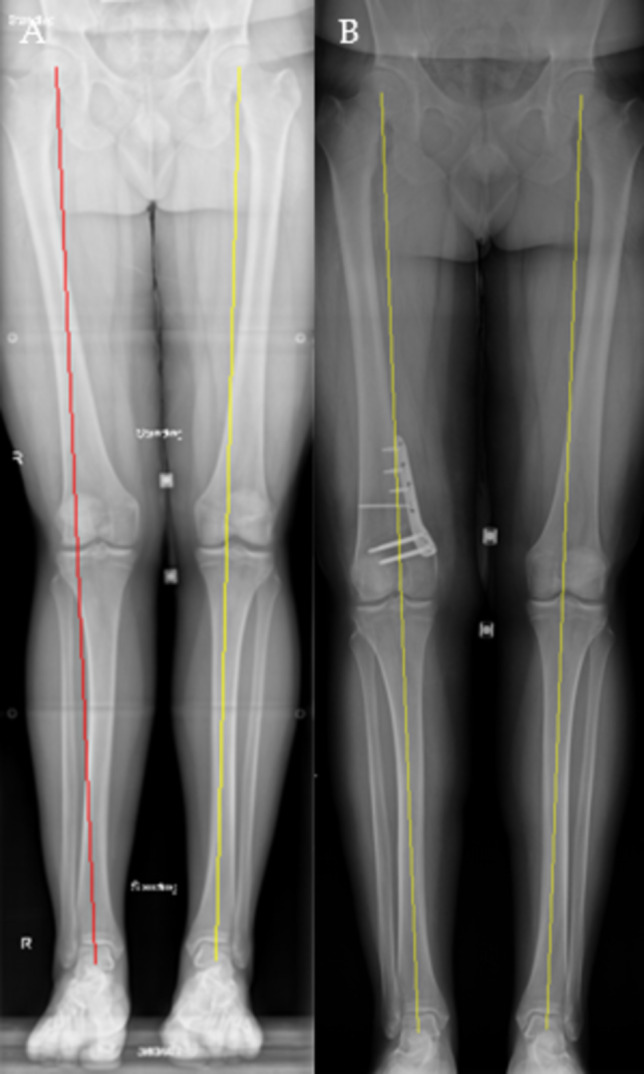
Purpose of review: Limb alignment correction about the knee joint is crucial for the protection of the meniscus, particularly in the setting of meniscal root repairs and meniscal allograft transplantation. Distal femoral osteotomies and high tibial osteotomies have been described to restore the anatomic alignment to aid in meniscal preservation. This article provides a review of knee alignment and biomechanics, various surgical interventions to correct knee malalignment, and the effect of malalignment on the treatment of meniscal pathology.
Recent findings: Both distal femoral and high tibial corrective osteotomies have been shown to slow the progression of osteoarthritis in the postoperative period. Moreover, corrective osteotomies have resulted in high patient satisfaction and good survival rates at mid- to long-term follow-up in patients with prior varus/valgus malalignment. Ongoing research is aimed to determine the best utilization for concomitant osteotomies in the setting and treatment of meniscal pathology with hopes of decreasing the progression of early-onset osteoarthritis, and ultimately, the conversion to total knee arthroplasty. Neutral alignment at the level of the knee joint results in optimal force distributions. Corrective valgus and varus osteotomies aim to restore neutral alignment with the goal of ligamentous and meniscal preservation, ideally slowing osteoarthritis progression.
Keywords: Distal femoral osteotomy; High tibial osteotomy; Meniscal allograft transplant; Root repair; Valgus malalignment; Varus malalignment; meniscus; distal femoral osteotomy; high tibial osteotomy; varus malalignment; valgus malalignment; osteoarthritis..
© 2025. The Author(s), under exclusive licence to Springer Science+Business Media, LLC, part of Springer Nature.
Conflict of interest statement
Declarations. Human and Animal Rights and Informed Consent: This article does not contain any studies with human or animal subjects performed by any of the authors. Competing Interest: Bruce A. Levy has received consulting fees from Arthrex; nonconsulting fees from Arthrex and Smith+Nephew; royalties from Arthrex; and has stock/stock options in COVR Medical.Andrew D. Carbone has received funding grants from Arthrex and Medical Device Business Services Inc; speaking and lecture fees from Arthrex, Smith+Nephew, Medwest Associates Inc, Micromed Inc, and Saxum Surgical Inc.Abhishek S. Kannan has received consulting fees from Linvatec Corporation; and speaking and lecture fees from Arthrex.
Figures


References
-
- Whiteside LA. Soft tissue balancing: the knee. J Arthroplasty. 2002;17(4 Suppl 1):23–7. 10.1054/arth.2002.33264. - PubMed
-
- Wang Y, Zeng Y, Dai K, Zhu Z, Xie L. Normal lower-extremity alignment parameters in healthy Southern Chinese adults as a guide in total knee arthroplasty. J Arthroplasty. 2010;25(4):563–70. 10.1016/j.arth.2009.03.021. - PubMed
-
- Micicoi G, Jacquet C, Sharma A, et al. Neutral alignment resulting from tibial vara and opposite femoral valgus is the main morphologic pattern in healthy middle-aged patients: an exploration of a 3D-CT database. Knee Surg Sports Traumatol Arthrosc. 2021;29(3):849–58. 10.1007/s00167-020-06030-4. - PubMed
-
- Uquillas C, Rossy W, Nathasingh CK, Strauss E, Jazrawi L, Gonzalez-Lomas G. Osteotomies about the knee: AAOS exhibit selection. J Bone Joint Surg Am. 2014;96(24):e199. 10.2106/JBJS.N.00270. - PubMed
-
- Fantini Pagani CH, Potthast W, Brüggemann GP. The effect of valgus bracing on the knee adduction moment during gait and running in male subjects with varus alignment. Clin Biomech (Bristol Avon). 2010;25(1):70–6. 10.1016/j.clinbiomech.2009.08.010. - PubMed
Publication types
LinkOut - more resources
Full Text Sources
Research Materials
