. 2025 Feb 21;44(1):64.
doi: 10.1186/s13046-025-03327-5.

The extracellular matrix protein type I collagen and fibronectin are regulated by β-arrestin-1/endothelin axis in human ovarian fibroblasts

Affiliations

The extracellular matrix protein type I collagen and fibronectin are regulated by β-arrestin-1/endothelin axis in human ovarian fibroblasts

Ilenia Masi et al. J Exp Clin Cancer Res. .

Abstract

Background: The invasive and metastatic spread of serous ovarian cancer (SOC) results from the cooperative interactions between cancer and stroma, which include extracellular matrix (ECM) and cellular components, including cancer-associated fibroblasts (CAFs). Soluble factors secreted by cancer and stromal cells contribute to stroma remodeling through the secretion of ECM proteins, providing a favorable environment for cancer cell dissemination. The peptide endothelin-1 (ET-1), through two G protein-coupled receptors (GPCR), endothelin receptor type A (ETAR) and B (ETBR), acts on both cancer and stromal cells, engaging the protein β-arrestin1 (β-arr1), to bolster SOC progression. However, its role in the regulation of the ECM proteins by ovarian fibroblasts is not understood. This study delves into the role of ET-1 as a regulator of type I collagen (Col1) and fibronectin (FN).

Methods: We used human primary ovarian fibroblasts (HOFs) and CAFs. The expression of Col1 (COL1A1) and FN (FN1) were detected by western blotting (WB), quantitative real time-polymerase chain reaction (qRT-PCR), immunofluorescence (IF), and confocal laser scanning microscopy (CLSM) in cells and tumor tissue sections from mice xenografts, while the transcription of COL1A1 was detected by luciferase reporter gene assay. The nuclear function of β-arr1 was evaluated by silencing and rescue expression with wild-type (WT) and nuclear mutant plasmid constructs, RNA seq and differential gene expression and gene sets enrichment analyses. The prognostic role of COL1A1, FN1, EDN1 (ET-1) and ARRB1 (β-arr1) gene expression was evaluated using the Kaplan-Meier plotter database and clinical ovarian cancer tissue samples.

Results: We demonstrated that ET-1 boosts Col1 and FN expression in HOFs, akin to ovarian CAF levels. Both receptors are implicated, evident from inhibitory effects after ETAR or ETBR antagonist treatments and notably with bosentan, a dual antagonist, in vitro and in vivo. At the molecular level, ET-1 triggers the activation of COL1A1 promoter activity and its enhanced expression via β-arr1 nuclear function. Transcriptome analysis of β-arr1-silenced HOFs confirms the nuclear role of β-arr1 in collagen and ECM remodeling-related protein transcriptional regulation. Accordingly, a high level of EDN1/ARRB1 expression in combination with either COL1A1 or FN1 is associated with the poor prognosis of SOC patients.

Conclusions: These findings hint at ET-1 involvement in ECM remodeling and early SOC stages by modulating the expression of Col1 and FN. Targeting ET-1 signaling with ETAR/ETBR antagonists might interfere with the ability of CAFs to produce key ECM proteins in this tumor.

Keywords: Endothelin receptors; Endothelin-1; Fibroblasts; Fibronectin; Ovarian cancer; Type I collagen; β-arrestin 1.


Conflict of interest statement

Declarations. Ethical approval: For in vivo animal studies, the experimental protocols complied with the principles of ARRIVE ( https://arriveguidelines.org ) according to institutional guidelines and the Italian Law (D-lgs 26/2014) and were approved by the National Ethics Committee for Animal Experimentation of the Italian Ministry of Health (authorization N1/2020- PR #365869604 Responsible Researcher Dr Laura Rosanò). Consent for publication: Not applicable. Competing interests: The authors declare no competing interests.

Figures

Fig. 1
Fig. 1
Kaplan-Meier analysis of overall survival (OS) or progression-free survival (PFS) curves in SOC patients with low or high FN1 or Col1A1 expression at stages 1 + 2 and 3 + 4
Fig. 2
Fig. 2
HOFs express ET-1, ETAR and ETBR, as well as COL1A1 and FN1. (A) Western Blotting (WB) analysis of indicated protein expression. Tubulin was used as a loading control. qRT-PCR analysis of (B)EDNRA (ETAR), EDNRB (ETBR), and EDN1 (ET-1) or (C)COL1A1 (Col1) and FN1 (Fibronectin) mRNA expression. (D-E) qRT-PCR analysis of COL1A1 and FN1 mRNA expression in HOFs stimulated with ET-1 (100 nM) at indicated time points and indicated as fold change compared to untreated cells. (F-G) Representative WB analysis of Col1 and FN expression in HOFs stimulated with ET-1 at the indicated time points. GAPDH was used as a loading control. Histograms, mean ± SD. n = 3. One-way ANOVA, n = 3 (*) p < 0.05, (**) p < 0.01, (***) p < 0.001
Fig. 3
Fig. 3
ET-1 induces Col1 and FN expression through ETAR and ETBR. Representative WB analysis of Col1 and FN protein expression in HOFs stimulated with ET-1 and/or Ambrisentan (AMB) (1 μm) and/or BQ788 (1 μm) and/or bosentan (BOS) (1 μm) for 24 h and 12 h, respectively. GAPDH was used as a loading control. Histograms, mean ± SD. One-way ANOVA (*) p < 0.05, (**) p < 0.01, (***) p < 0.001, (****) p < 0.0001
Fig. 4
Fig. 4
ET-1 induces the expression of COL1A1 and FN1 through ETA/BR in ovarian CAFs. (A) qRT-PCR analysis of COL1A1 mRNA expression in CAFs stimulated with ET-1 (100 nM) at indicated time points and indicated as fold change compared to untreated cells. One-way ANOVA (*) p < 0.05, (**) p < 0.01, (***) p < 0.001, (****) p < 0.0001. (B) Representative WB analysis of Col1 and FN protein expression in HOFs and CAFs stimulated with ET-1 for 24 h. GAPDH was used as a loading control. n = 2 (C) Representative WB analysis of Col1 and FN protein expression in CAFs grown with serum-free medium (CTR) in the presence or absence of AMB or BQ788 or BOS (24 h). GAPDH was used as a loading control. Histograms, mean ± SD. One-way ANOVA, n = 3. (*) p < 0.05, (**) p < 0.01, (***) p < 0.001), (****) p < 0.0001
Fig. 5
Fig. 5
ET-1/ETA/ETBR induces expression of COL1A1 through nuclear β-arr1. (A) qRT-PCR analysis of COL1A1 mRNA expression in HOFs silenced for β-arr1 (si-ARRB1) and transfected with Mock or ARRB1-Q394L-FLAG or ARRB1-WT-FLAG and stimulated with ET-1 (24 h) and shown as fold over CTR. (B) Representative WB of whole cell lysates from HOFs transfected as in A. and probed with Abs to β-arr1 and FLAG. GAPDH was used as loading control. (C)COL1A1 promoter activity in HOFs treated as in A. or (D) in HOFs stimulated or not with ET-1 and/or BOS (24 h), calculated as Firefly Luc value/GAL enzyme activity. Histograms, mean ± SD. One-way ANOVA, n = 3. (*) p < 0.05, (**) p < 0.01, (***) p < 0.001, (****) p < 0.0001. (E) IF analysis of HOFs stimulated or not with ET-1 and/or BOS (24 h). Cells were stained for Col1 (green), F-actin (red), and DAPI (blue). Scale bar 50 μm. Histograms, mean ± SD. One-way ANOVA, n = 3. (*) p < 0.05
Fig. 6
Fig. 6
RNA-seq analysis of HOFs silenced or not for β-arr1. (A) Volcano plot showing genes’ −log10(FDR) and log2(fold change). Significantly modulated genes (FDR<0.05 and|log2 fold change| > 1) are highlighted in red, whereas not significantly modulated genes are indicated in dark blue. The vertical dashed lines indicate the thresholds for up- and down-regulated genes and the horizontal dashed line represents the FDR threshold. The top 50 most significant DEGs are labeled. (B) Dot plot of over-representation analysis of upregulated and downregulated DEGs which shows significantly enriched gene sets according to GO CC terms. Dot size indicates a ratio between the number of genes participating in the current GO CC category and the number of up or downregulated DEGs. The dot color indicates the FDR (qvalue) associated with each enriched term. (C) Heatmap of z-score scaled expression values of genes belongs to the collagen-containing extracellular matrix category. (D) Analysis of ARRB1 expression in 7 cancer stroma and 7 matched cancer epithelia from 18 ovarian cancer patients. One-way ANOVA
Fig. 7
Fig. 7
ET-1 receptor blockade inhibits the expression of Col1 and FN in SOC/HOF xenografts. (A) CLSM examinations (3D reconstruction images) of FFPE tissue Sect. (5 μm thick) from untreated (CTR) and AMB or BOS-treated mice. Sections were stained for FN detection (red) in combination with anti-αSMA (green, left panels) or anti-Col1 primary Ab, shown in green (right panels). Nuclei were stained with DAPI (blue). Separate channels and merged images are reported. Scale bar, 50 μm. n = 2. (B) Representative WB of whole cell lysates from metastatic nodules probed with Abs to Col1 and FN. Histograms, mean ± SD. One-way ANOVA, n = 3. (*) p < 0.05, (**) p < 0.01, (***) p < 0.001, (****) p < 0.0001
Fig. 8
Fig. 8
High EDN1/ARRB1/COL1A1 and EDN1/ARRB1/FN1 expression correlates with poor prognosis of SOC patients. Kaplan-Meier analysis of overall survival (OS) or progression-free survival (PFS) curves in SOC patients with low or high EDN1/ARRB1(A) or EDN1/ARRB1/COL1A1(B) or EDN1/ARRB1/FN1(C) expression at stage 1 + 2 and 3 + 4
Fig. 9
Fig. 9
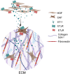
Schematic representation of proposed mechanism by which ET-1 promotes ECM protein expression and production through ETAR/ETBR axis in ovarian fibroblasts. Autocrine and paracrine ET-1 activates ETAR and ETBR in HOFs and CAFs, leading to Col1 and FN release in ovarian ECM. Created with BioRender.com (Agreement Number IK273AZ978)

References

    1. Kai F, Drain AP, Weaver VM. The Extracellular Matrix modulates the metastatic journey. Dev Cell. 2019;49:332–46. - PMC - PubMed
    1. Anderson NM, Simon MC. The tumor microenvironment. Curr Biol. 2020;30:R921–5. - PMC - PubMed
    1. Davenport AP, Hyndman KA, Dhaun N, Southan C, Kohan DE, Pollock JS, et al. Endothelin Pharmacol Rev. 2016;68:357–418. - PMC - PubMed
    1. Rosanò L, Spinella F, Bagnato A. Endothelin 1 in cancer: biological implications and therapeutic opportunities. Nat Rev Cancer. 2013;13:637–51. - PubMed
    1. Salani D, Di Castro V, Nicotra MR, Rosanò L, Tecce R, Venuti, et al. Role of endothelin-1 in neovascularization of ovarian carcinoma. Am J Pathol. 2000;157:1537–47. - PMC - PubMed

MeSH terms

LinkOut - more resources