. 2025 Feb 21;13(1):39.
doi: 10.1186/s40478-025-01941-0.

Neuronal TDP-43 aggregation drives changes in microglial morphology prior to immunophenotype in amyotrophic lateral sclerosis

Affiliations

Neuronal TDP-43 aggregation drives changes in microglial morphology prior to immunophenotype in amyotrophic lateral sclerosis

Molly E V Swanson et al. Acta Neuropathol Commun. .

Abstract

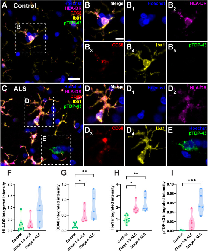
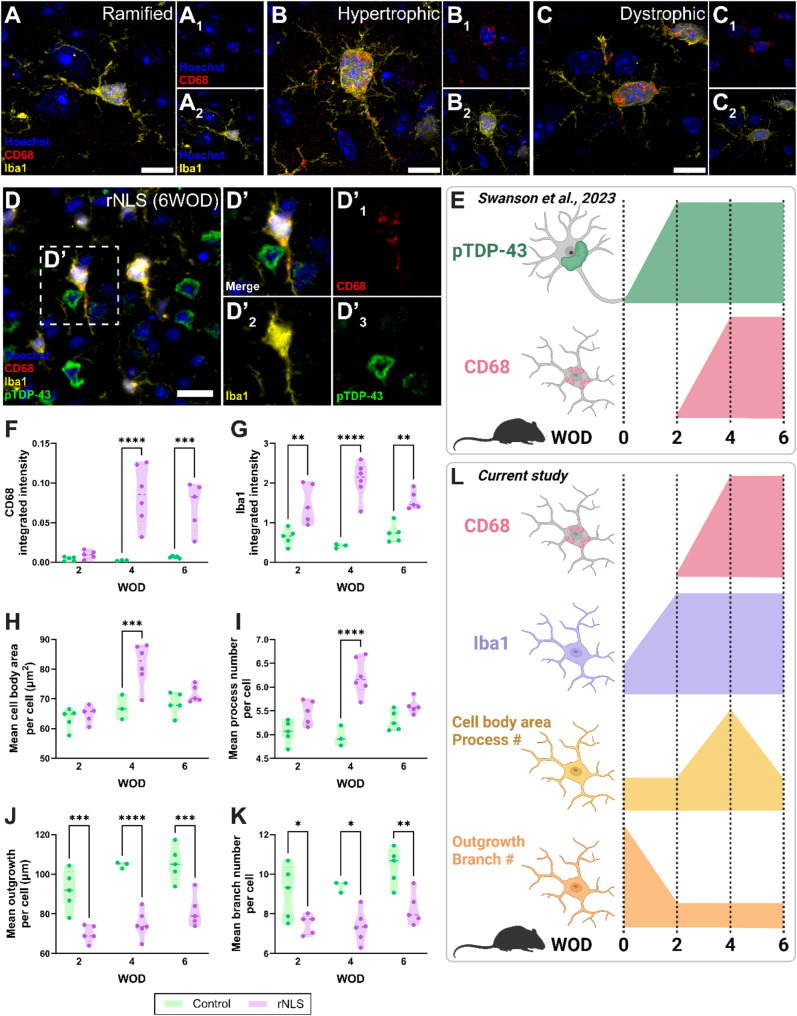
Microglia are the innate immune cells of the brain with the capacity to react to damage or disease. Microglial reactions can be characterised in post-mortem tissues by assessing their pattern of protein expression, or immunophenotypes, and cell morphologies. We recently demonstrated that microglia have a phagocytic immunophenotype in early-stage ALS but transition to a dysfunctional immunophenotype by end stage, and that these states are driven by TAR DNA-binding protein 43 (TDP-43) aggregation in the human brain. However, it remains unclear how microglial morphologies are changed in ALS. Here we examine the relationship between microglial immunophenotypes and morphologies, and TDP-43 pathology in motor cortex tissue from people with ALS and from a TDP-43-driven ALS mouse model. Post-mortem human brain tissue from 10 control and 10 ALS cases was analysed alongside brain tissue from the bigenic NEFH-tTA/tetO-hTDP-43∆NLS (rNLS) mouse model of ALS at distinct disease stages. Sections were immunohistochemically labelled for microglial markers (HLA-DR, CD68, and Iba1) and phosphorylated TDP-43 (pTDP-43). Single-cell microglial HLA-DR, CD68, and Iba1 average intensities, and morphological features (cell body area, process number, total outgrowth, and branch number) were measured using custom image analysis pipelines. In human ALS motor cortex, we identified a significant change in microglial morphologies from ramified to hypertrophic, which was associated with increased Iba1 and CD68 levels. In the rNLS mouse motor cortex, the microglial morphologies changed from ramified to hypertrophic and increased Iba1 levels occurred in parallel with pTDP-43 aggregation, prior to increases in CD68 levels. Overall, the evidence presented in this study demonstrates that microglia change their morphologies prior to immunophenotype changes. These morphological changes may prime microglia near neurons with pTDP-43 aggregation for phagocytosis, in turn triggering immunophenotype changes; first, to a phagocytic state then to a dysfunctional one.

Keywords: Amyotrophic lateral sclerosis; CD68; Dystrophic; Hypertrophic; Iba1; Microglia; Ramified; TDP-43.


Conflict of interest statement

Declarations. Ethics approval and consent to participate: All human brain tissue was donated to the Neurological Foundation Human Brain Bank following donor and donor family consent, and its use in this study was approved by the University of Auckland Human Participants Ethics committee (protocol number 011654). Animal ethics approval was obtained from The University of Queensland (#QBI/131/18), and experiments were conducted in accordance with the Australian code of practice for the care and use of animals for scientific purposes. Consent for publication: Not applicable. Competing interests: The authors declare no competing interests.

Figures

Fig. 1
Fig. 1
Increased microglial CD68 and Iba1 levels and pTDP-43 abundance in the human ALS motor cortex. Immunohistochemical labelling was used to visualise microglia and pTDP-43 in the motor cortex from human control (A-B), stage 1–3 (not shown), and stage 4 ALS (C-E) cases. Microglial markers, HLA-DR (magenta), CD68 (red), and Iba1 (yellow), were co-labelled with pTDP-43 (green) and a Hoechst nuclear counterstain (blue). Scale bar (A and C) = 20 μm and scale bar (B, D, and E) = 10 μm. Total microglia were identified by creating separate binary masks from thresholded HLA-DR, CD68, and Iba1 images which were then combined to create a microglial master mask [39, 69, 86]. The total integrated intensities of HLA-DR (F), CD68 (G), and Iba1 (H) were measured within this master mask, normalised to tissue area and compared between control, stage 1–3 ALS, and stage 4 ALS cases. The total integrated intensity of pTDP-43 aggregates normalised to tissue area was quantified and compared between normal, stage 1–3 ALS, and stage 4 ALS cases (I). Data presented as truncated violin plots with median and quartiles shown; control n = 10, stage 1–3 ALS n = 5, and stage 4 ALS n = 5. All intensity measures were compared between case groups with multiple Mann-Whitney tests and multiple comparisons were controlled for using a False Discovery Rate of 0.01, as determined by the two-stage step-up method of Benjamini, Krieger, and Yekutieli. Significances of difference between case groups: *p ≤ 0.05, **p ≤ 0.01, ***p ≤ 0.001
Fig. 2
Fig. 2
Microglial morphologies change in human ALS and correlate with CD68 and Iba1 levels. Microglial cell bodies and processes were identified using Iba1 immunoreactivity (A, yellow) and used to quantify morphology measures per cell (A, white): cell body area (B), process number (C), outgrowth (D), and branch number (E). Single-cell measures were averaged across all microglia in each case to give a mean measure per cell which was compared between control, stage 1–3 ALS, and stage 4 ALS cases. Data presented as truncated violin plots with median and quartiles shown; control n = 10, stage 1–3 ALS n = 5, and stage 4 ALS n = 5. All morphology measures were compared between case groups with multiple Mann-Whitney tests and multiple comparisons were controlled for using a False Discovery Rate of 0.01, as determined by the two-stage step-up method of Benjamini, Krieger, and Yekutieli. Significances of difference between case groups: *p ≤ 0.05. Measures of pTDP-43 pathology, microglial marker intensities, and microglial morphology were sequentially correlated in all cases (n = 20) using Spearman correlations (F). The resulting r value from each correlation is presented in the correlation matrix and colour coded relative to strength. When r ≤ -0.7 or r ≥ 0.7 and p ≤ 0.05, correlations were considered statistically significant and strong. When − 0.7 < r ≤ -0.4 or 0.7 > r ≥ 0.4 and p ≤ 0.05, correlations were considered statistically significant and moderate
Fig. 3
Fig. 3
Microglial clusters enriched in human ALS are characterised by high cell body area, low branch number, and high Iba1 and CD68 levels. Louvain clustering was carried out using the single-cell average intensities of CD68, HLA-DR, and Iba1, and morphological measures (cell body area, process number, outgrowth, and branch number) from a randomly subsampled 7,340 microglia per case group (22,020 cells total), resulting in 11 unique clusters (arbitrarily numbered) (A). The percentage of microglia in each cluster was compared between control, stage 1–3 ALS, and stage 4 ALS cases using a 2-way ANOVA with Tukey’s multiple comparisons test; data presented as truncated violin plots with median and quartiles shown; control n = 10, stage 1–3 ALS n = 5, and stage 4 ALS n = 5. For clusters depleted (1 and 8; B) and enriched (4 and 10; C) in ALS cases, the percentage of microglia in each cluster was correlated with pTDP-43 integrated intensity (I.I.) using Spearman’s correlation; each correlation was carried out using a total n = 10 cases, with stage 1–3 ALS and stage 4 ALS cases colour-coded and r and p values presented for each correlation. The mean single-cell CD68 and Iba1 average intensity was compared between clusters using a Kruskal-Wallis test with Dunn’s multiple comparisons test (D and E); data presented for ALS-depleted and ALS-enriched clusters as a box and whisker graph, with minimum, maximum, and median shown (n = 20). The mean cell body area and single-cell branch number were determined for all clusters (F); data presented for ALS-depleted and ALS-enriched clusters as a box and whisker graph, with minimum, maximum, and median shown (n = 20). Significance of differences between groups: *p ≤ 0.05, **p ≤ 0.01, ***p ≤ 0.001, ****p ≤ 0.0001. Confocal microscopy was used to visualise ramified (G), hypertrophic (H), and dystrophic microglial morphologies (I); example images from stage 4 ALS case, MN29, are shown; scale bars = 10 μm
Fig. 4
Fig. 4
Microglial Iba1 and morphological changes occur parallel to pTDP-43 aggregation and prior to CD68 changes in the TDP-43-driven rNLS mouse model of ALS. Immunohistochemical labelling of Iba1 and CD68 was carried out on motor cortex tissue from bigenic NEFH-tTA/tetO-hTDP-43∆NLS (rNLS) and single transgenic tetO-hTDP-43∆NLS (control) mice at 2, 4, and 6 weeks off DOX (WOD). Confocal microscopy was used to visualise ramified (A), hypertrophic (B), and dystrophic microglial morphologies; example images from rNLS at 6 WOD are shown; Scale bars = 10 μm. Images previously analysed in Swanson et al., (2023) were reanalysed to investigate temporal changes in microglial morphology measures relative to pTDP-43 aggregation; an example immunohistochemical image of CD68 (red), and Iba1 (yellow), pTDP-43 (green) and a Hoechst nuclear counterstain (blue) labelling from a rNLS mouse at 6 WOD is shown (D); scalebar = 20 μm. Summary of Swanson et al., (2023) findings is shown in E; created with BioRender.com. Total microglia were identified by creating separate binary masks from CD68 and Iba1 images which were then combined to create a microglial master mask. The integrated intensities of CD68 (F) and Iba1 (G) were measured within this master mask and compared between control and rNLS mice at 2, 4, and 6 WOD. Microglial cell bodies and processes were identified using Iba1 immunoreactivity and used to quantify morphology measures per cell: cell body area (H), process number (I), outgrowth (J), and branch number (K). Single-cell measures were averaged across all microglia in each case to give a mean measure per cell which was compared between control and rNLS mice at 2, 4, and 6 WOD. Data presented as truncated violin plots with median and quartiles shown; control n = 3–5, rNLS n = 4–6. Each intensity and morphology measure was compared between case groups at each WOD timepoint with 2-way ANOVA with Tukey’s multiple comparisons test. Significance of differences between case groups: *p ≤ 0.05, **p ≤ 0.01, ***p ≤ 0.001, ****p ≤ 0.0001. Summary of current study’s findings is shown in L; created with BioRender.com

References

    1. Aderem A, Underhill DM (2002) Mechanisms of Phagocytosis in macrophages. Annu Rev Immunol 17:593–623 - PubMed
    1. Gibbons HM, Smith AM, Teoh HH, Bergin PM, Mee EW, Faull RLM et al (2011) Valproic acid induces microglial dysfunction, not apoptosis, in human glial cultures. Neurobiol Dis 41:96–103 - PubMed
    1. Rogers J, Strohmeyer R, Kovelowski CJ, Li R (2002) Microglia and inflammatory mechanisms in the clearance of amyloid beta peptide. Glia 40:260–269 - PubMed
    1. Simard AR, Soulet D, Gowing G, Julien J-P, Rivest S (2006) Bone marrow-derived microglia play a critical role in restricting senile plaque formation in Alzheimer’s disease. Neuron 49:489–502 - PubMed
    1. Chhor V, Le Charpentier T, Lebon S, Oré M-V, Celador IL, Josserand J et al (2013) Characterization of phenotype markers and neuronotoxic potential of polarised primary microglia in vitro. Brain Behav Immun 32:70–85 - PMC - PubMed

Publication types

MeSH terms