. 2025 Apr;12(15):e2410438.
doi: 10.1002/advs.202410438. Epub 2025 Feb 22.

An Anti-CD147 Antibody-Drug Conjugate Mehozumab-DM1 is Efficacious Against Hepatocellular Carcinoma in Cynomolgus Monkey

Affiliations

An Anti-CD147 Antibody-Drug Conjugate Mehozumab-DM1 is Efficacious Against Hepatocellular Carcinoma in Cynomolgus Monkey

Wan Huang et al. Adv Sci (Weinh). 2025 Apr.

Abstract

Effective treatment strategies are urgently needed for hepatocellular carcinoma (HCC) patients due to frequent therapeutic resistance and recurrence. Antibody-drug conjugate (ADC) is a specific antibody-drug conjugated with small molecular compounds, which has potent killing activity against cancer cells. However, few ADC candidates for HCC are undergoing clinical evaluation. CD147 is a tumor-associated antigen that is highly expressed on the surface of tumor cells. Here CD147 is found significantly upregulated in tumor tissues of HCC. Mehozumab-DM1, a humanized anti-CD147 monoclonal antibody conjugated with Mertansine (DM1) is developed. Mehozumab-DM1 is effectively internalized by cancer cells and demonstrated potent antitumor efficacy in HCC cells. In vivo evaluation of Mehozumab-DM1 is conducted in a CRISPR-mediated PTEN and TP53 mutation cynomolgus monkey liver cancer model, which is poorly responsive to sorafenib treatment. Mehozumab-DM1 demonstrated potent tumor inhibitory efficacy at doses of 0.2 and 1.0 mg kg-1 treatment groups in cynomolgus monkey. No treatment-related adverse reactions or body weight loss are observed. Interestingly, Mehozumab-DM1 treatment induced RIPK-dependent tumor cell necroptosis through inhibiting IκB kinase/NF-κB pathway. In conclusion, Mehozumab-DM1 potently inhibits hepatoma through effective internalization to release payload and inducing cell necroptosis to enhance the bystander effect, which is a promising treatment for refractory HCC.

Keywords: antibody‐drug conjugate; cell necroptosis; hepatocellular carcinoma; therapeutic resistance.


Conflict of interest statement

The authors declare no conflict of interest

Figures

Figure 1
Figure 1
Expression of CD147 was significantly upregulated in tumor tissues of HCC and correlated with targeted therapy resistance. A) CD147 protein levels were quantitatively compared in tumor and normal tissues of HCC patients based on the CPTAC dataset(n = 153). Representative image of IHC of CD147 in para‐tumor and tumor tissues from patients with HCC. Scale bars represent 50 µm. B) Transcription level of CD147 (BSG) in the normal tissue, tumor tissue, different stages of HCC according to AJCC staging, and different pathological grades of HCC. Data from the TCGA dataset. C) Gene expression of CD147 (BSG) was quantitatively compared in the tumor tissues from trastuzumab (n = 45), bevacizumab (n = 12) and sorafenib (n = 17) non‐response patients and response patients based on CTR‐DB dataset. D) BSG was quantitatively compared in sorafenib‐resistant HepG2 cells and HepG2 cells based on the GEO dataset. Independent Student's t‐test was used to compare the mean expression level of two different groups. *p < 0.05, **p < 0.01, ***p < 0.001. NS, is not significant.
Figure 2
Figure 2
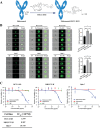
Mehozumab‐DM1 is effectively internalized and exhibits antitumor efficacy in HCC cells. A) Diagram of the preparation process of Mehozumab‐DM1. Mehozumab was conjugated to DM1 via the non‐cleavable linker SMCC, forming Mehozumab‐DM1, with an average drug‐to‐antibody ratio (DAR) of 5.80. B) Representative imaging flow cytometry images showing cellular internalization of IgG, Mehozumab, and Mehozumab‐DM1 labeled with FITC (green) in HCC‐LM3 and Huh‐7 HCC cell lines (left). Signal intensity analysis for Mehozumab‐DM1 antibody‐mediated cell internalization (right). Independent Student's t test was used to compare the mean expression level of two different groups. *p < 0.05, **p < 0.01, ***p < 0.001. ns, not significant. BF, bright field; IgG, immunoglobulin G. C) Cell Counting Kit‐8 experiments were conducted to evaluate the inhibitory efficacy of Mehozumab‐DM1 on hepatocellular carcinoma cell lines HCC‐LM3, MHCC97‐H, and Huh‐7. Mehozumab‐DM1, Mehozumab, and IgG with different concentration gradients were added to the cells and treated for 72 h. IC50 values for three cell lines were calculated in the table.
Figure 3
Figure 3
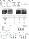
In vivo inhibitory efficacy of Mehozumab‐DM1 in CRISPR‐mediated cynomolgus monkey liver cancer model. A) Correlation of TP53 (left) and PTEN (right) mutation and IC50 of sorafenib in HCC cell lines (data from The Genomics of Drug Sensitivity in Cancer Project, TP53‐mut: n = 10, WT: n = 5. PTEN‐mut: n = 1 WT: n = 14). B) Schematic design of in vivo study in CRISPR‐mediated cynomolgus monkey liver cancer model. C) Typical photos of ultrasonic scanning before and after ADC treatment for 8 weeks in Mehozumab‐DM1 intravenous injection 0.2 and 1.0 mg kg−1 group. The detailed data of tumor volume before and after treatment of each cynomolgus monkey, and tumor volume inhibition rate was shown in the table below. D) Kaplan Meier curves and log‐rank analyses to investigate the overall survival in cynomolgus monkey treatment with physiological saline, 0.2 and 1 mg kg−1 ADC. E) The level of AFP in peripheral blood of the cynomolgus monkey before and after the ADC treatment for 4 and 8 weeks in control and Mehozumab‐DM1 treatment 0.2 and 1.0 mg kg−1 groups. F) The body weight of the cynomolgus monkey was recorded for 10 weeks during ADC treatment. G,H) The level of AST and ALT in peripheral blood of the cynomolgus monkey before and after the ADC treatment for 4 and 8 weeks in control and Mehozumab‐DM1treatment groups. Independent Student's t test was used to compare the mean expression level of two different groups. *p < 0.05, **p < 0.01, ns, not significant. AFP: alpha‐fetoprotein; AST: Aspartate aminotransferase; ALT: Alanine aminotransferase.
Figure 4
Figure 4
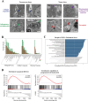
Cell necroptosis induced by Mehozumab‐DM1 in tumor tissues from cynomolgus monkey. A) Pathological changes in tumor tissues and precancerous tissues of cynomolgus monkeys after ADC treatment were detected by electron microscopy. ADC treatment led to obvious collagen deposition (blue arrow) and inflammatory cell infiltration (green arrow) in precancerous tissue (left). In tumor tissue (right), mitochondrial swelling and ER expansion in the cell (purple arrow), and typical cell necroptosis (red arrow) were observed. B) Total RNA from the tumor tissue of physiological saline control cynomolgus monkey and cynomolgus monkey treated with 1mg kg−1 ADC was extracted to carry out RNA‐seq. The functional interpretation of dysregulated RNA was annotated using GO analysis. C) Differential genes between control and ADC treatment cynomolgus monkey were enriched by Gene Set Enrichment Analysis (GSEA). Genes related to the intrinsic apoptotic and cell death signaling pathway rank high in their GSEA enrichment scores. D) GSEA to identify the apoptosis and programmed cell death signaling pathways enriched in differential genes between control and ADC treatment cynomolgus monkey.
Figure 5
Figure 5
Mehozumab‐DM1 treatment induced RIPK‐dependent tumor cell necroptosis through inhibiting IκB kinase / NF‐κB pathway. A) Annexin V/7‐AAD apoptosis analysis by flow cytometry in MHCC97‐H cells (left) and Huh‐7 cells (right) after 30 and 60 µg mL−1 ADC treatment for 72 h. Independent Student's t test was used to compare the mean expression level of two different groups. *p < 0.05, **p < 0.01, *** p < 0.001, ns, not significant. B) Immunoblotting analysis to detect the necroptosis markers RIPK3 and MLKL in Huh‐7 cells, after 24 and 48 h Mehozumab‐DM1 treatment. C) GSEA enrichment analysis of NF‐κB pathway in differential genes between control and ADC treatment cynomolgus monkey. D) Immunoblotting analysis of TNFAIP3/A20 (tumor necrosis factor alpha‐induced protein 3), phosphorylated IKK (inhibitor of kappa B kinase), and P65 (nuclear factor kappa B p65) after 24 and 48 h Mehozumab‐DM1 treatment. E) Diagram to summarize the mechanisms that Mehozumab‐DM1 inhibits hepatoma growth in HCC patients that are resistant to targeted therapy. Mehozumab‐DM1 was effectively internalized by cancer cells to release the payload, thereby inhibiting microtubule assembly and cell mitosis. Besides, Mehozumab‐DM1 induced RIPK‐dependent cell necroptosis by inhibiting the IκB kinase/NF‐κB pathway caused by inhibiting CD147‐induced changes in programmed cell death signaling. Furthermore, cell necroptosis causes cell membrane damage, the free payload was released outside the cell, thereby enhancing the bystander effect of ADC.

References

    1. Liu C., Zhou C., Xia W., Zhou Y., Qiu Y., Weng J., Zhou Q., Chen W., Wang Y. N., Lee H. H., Wang S. C., Kuang M., Yu D., Ren N., Hung M. C., Nat. Commun. 2024, 15, 1009. - PMC - PubMed
    1. Ducreux M., Abou‐Alfa G. K., Bekaii‐Saab T., Berlin J., Cervantes A., de Baere T., Eng C., Galle P., Gill S., Gruenberger T., Haustermans K., Lamarca A., Laurent‐Puig P., Llovet J. M., Lordick F., Macarulla T., Mukherji D., Muro K., Obermannova R., O'Connor J. M., O'Reilly E. M., Osterlund P., Philip P., Prager G., Ruiz‐Garcia E., Sangro B., Seufferlein T., Tabernero J., Verslype C., Wasan H., et al., ESMO Open 2023, 8, 101567. - PMC - PubMed
    1. Rumgay H., Arnold M., Ferlay J., Lesi O., Cabasag C. J., Vignat J., Laversanne M., McGlynn K. A., Soerjomataram I., J. Hepatol. 2022, 77, 1598. - PMC - PubMed
    1. Yang X., Yang C., Zhang S., Geng H., Zhu A. X., Bernards R., Qin W., Fan J., Wang C., Gao Q., Cancer Cell 2024, 42, 180. - PubMed
    1. Khongorzul P., Ling C. J., Khan F. U., Ihsan A. U., Zhang J., Mol. Cancer Res. 2020, 18, 3. - PubMed

MeSH terms

LinkOut - more resources