. 2025 Apr;12(15):e2412881.
doi: 10.1002/advs.202412881. Epub 2025 Feb 22.

CpG-Based Nanovaccines Enhance Ovarian Cancer Immune Response by Gbp2-Mediated Remodeling of Tumor-Associated Macrophages

Affiliations

CpG-Based Nanovaccines Enhance Ovarian Cancer Immune Response by Gbp2-Mediated Remodeling of Tumor-Associated Macrophages

Jiaqiang Xiong et al. Adv Sci (Weinh). 2025 Apr.

Abstract

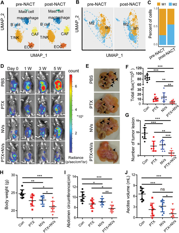
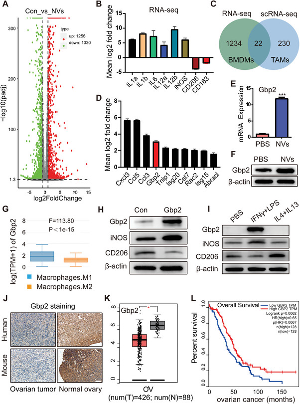
CpG oligodeoxynucleotides (CpG), as an immunoadjuvant, can facilitate the transformation of tumor-associated macrophages (TAMs)into tumoricidal M1 macrophages. However, the accumulation of free CpG in tumor tissues remains a substantial challenge. To address this, a nanovaccine (PLGA-CpG@ID8-M) is engineered by encapsulating CpG within PLGA using ID8 ovarian cancer cell membranes (ID8-M). This nanovaccine demonstrates remarkable efficacy in reprogramming TAMs in ovarian cancer and significantly extends survival in ID8-bearing mice. Notably, these findings indicate that the nanovaccine can also mitigate chemotherapy-induced immunosuppression by increasing the proportion of M1-like TAMs and reducing the expression of CD47 on tumor cells, thereby achieving a synergistic effect in tumor immunotherapy. Mechanistically, through transcriptome sequencing (RNA-seq), single-cell RNA sequencing (scRNA-seq), and mass spectrometry-based proteomics, it is elucidated that the nanovaccine enhances the expression of Gbp2 and promotes the recruitment of Pin1, which activates the NFκB signaling pathway, leading to the M1 polarization of TAMs. Furthermore, macrophages with elevated Gbp2 expression significantly inhibit tumor growth in both ID8 ovarian cancer and 4T1 breast cancer models. Conversely, targeting Gbp2 diminishes the antitumor efficacy of the nanovaccine in vivo. This study offers an innovative approach to immunotherapy and elucidates a novel mechanism (Gbp2-Pin1-NFκB pathway) for remodeling TAMs.

Keywords: Gbp2; TAMs; immunotherapy; nanovaccine; ovarian cancer.


Conflict of interest statement

The authors declare no conflict of interest.

Figures

Figure 1
Figure 1
Synthesis and safety evaluation of nanovaccines. A) Schematic illustration of the synthesis of PLGA‐CpG@ID8‐M nanovaccine. B) Size distribution and TEM images of PLGA‐CpG@ID8‐M (Scale bar: 100 nm). C) Surface zeta potential measurements of PLGA‐CpG, CpG, ID8‐M, and PLGA‐CpG@ID8‐M. D) Long‐term stability of PLGA‐CpG@ID8‐M in PBS and 10% FBS. E) SDS‐PAGE protein analysis of PLGA‐CpG, CpG, ID8‐M, and PLGA‐CpG@ID8‐M using Coomassie blue staining. F) Cell viability of RAW264.7 macrophages incubated with various concentrations of PLGA‐CpG@ID8‐M for 24 h. G) Secretion of IFN‐γ detected by ELISA after BMDMs were cocultured with the nanovaccine for 24 h. H) CLSM images of ID8 and RAW264.7 cells incubated with DiI‐labeled PLGA‐CpG@ID8‐M for 4 h, scale bars: 20 µm. I,J) Tumor‐targeting capability of the nanovaccine (1 nmol of CpG per mouse) assessed by IVIS following intravenous injection into ID8 tumor‐bearing mice for 6 h (n = 3). Data are presented as mean ± SD, analyzed using an unpaired two‐sided Student's t test (*p < 0.05, **p < 0.01, ***p < 0.001).
Figure 2
Figure 2
Nanovaccines induce effective antitumor immune responses. A,B) mRNA expression levels of canonical M1 and M2 macrophage biomarkers in BMDMs treated with nanovaccines for 12 h (n = 3). C–E) The secretion of proinflammatory cytokines IFN‐γ, TNF‐α, and IL‐6 was measured by ELISA following a 12 h treatment of BMDMs with nanovaccines (n = 3). F,G) Expression of CD80 and CD206 in BMDMs were assessed by flow cytometry following 12 h treatment with nanovaccines (n = 3). H,I) Expression of iNOS was detected by immunofluorescence following nanovaccine treatment for 12 h (n = 3), scale bar: 50 µm. J,K) Expression of iNOS and Arg1 was detected by western blot following nanovaccine treatment for 12 h (n = 3). L,M) Expression of HIF1A and CD47 was detected by RT‐PCR and western blot following nanovaccine treatment for 12 h (n = 3). N) Phagocytic activity of BMDMs on ID8 cells following nanovaccine treatment for 24 h, scale bar: 50 µm. O) Fold change in BMDM‐mediated phagocytosis of ID8 cells over 72 h (n = 3). P) Schematic representation of the synergistic antitumor effects of nanovaccines. Data are presented as mean ± SD, analyzed using an unpaired two‐sided Student's t‐test (*p < 0.05, **p < 0.01, ***p < 0.001).
Figure 3
Figure 3
Nanovaccine combined with chemotherapy achieves synergistic antitumor effects in vivo. A) UMAP (uniform manifold approximation and projection) plot depicting all cells from prospective tumor samples of 11 patients with HGSOC before and after NACT (data from GSE165897). B,C) Classification of M1 and M2 phenotypes within the macrophage subpopulations. D,F) antitumor effects of PBS, PTX (20 mg kg−1, intraperitoneal injection), NVs (CpG content at 1 nmol per mouse, intravenous injection), and the combination (NVs + PTX) observed at various time points using IVIS in mouse models of ovarian cancer with intraperitoneal metastasis (n = 8). E,G) Assessment of the number of abdominal tumor lesions observed at the fifth week across different groups (n = 8). H–J) Analysis of body weight, abdominal circumference, and ascites volume at the fifth week across different groups (n = 8). Data are presented as mean ± SD, analyzed using an unpaired two‐sided Student's t test (ns: not significant, *p < 0.05, **p < 0.01, ***p < 0.001).
Figure 4
Figure 4
Nanovaccines induce antitumor effects in ovarian cancer by improving the immune microenvironment. A) Schematic illustration of the treatment approach for subcutaneous ovarian cancer tumor models. B–D) Changes in tumor size, volume, and weight of ID8 ovarian cancer following different treatments as indicated (n = 8). E) Survival curves of mice in different groups observed over a continuous 60 d period (n = 7). F–H) Proportions of M2/M1 macrophages (CD206/CD80) in different groups of ID8 tumor tissues, assessed by multiplex immunohistochemistry and flow cytometry (n = 4), scale bar: 20 µm. I,J) Proportion of CD8a+ T cells in different groups of ID8 tumor tissues, determined by flow cytometry (n = 4). K,L) Immunohistochemical staining and statistical analysis of CD47 in the different groups, scale bar: 50 µm. Data are presented as mean ± SD, analyzed using an unpaired two‐sided Student's t test (ns: not significant, *p < 0.05, **p < 0.01, ***p < 0.001).
Figure 5
Figure 5
The role of nanovaccine‐induced Gbp2 in tumor prognosis. A) Number of differentially expressed genes identified in the RNA‐seq analysis. B) RNA‐seq results showing the expression levels of IL1a, IL1b, IL6, IL12a, IL12b, CD206, and CD163 in BMDMs treated with nanovaccines for 12 h. C,D) Enrichment of differentially expressed genes identified in both RNA‐seq and scRNA‐seq datasets. E,F) Detection of Gbp2 expression following BMDM activation with nanovaccines, measured by RT‐PCR and western blot. G) Expression levels of Gbp2 in M1 and M2 macrophages, obtained from the GEPIA database. H,I) Immunoblot (IB) analysis of the indicated proteins derived from RAW264.7 macrophages stably overexpressing Gbp2 (H) and those induced by (LPS + IFN‐γ) and (IL4 + IL13) (I). J) Immunohistochemical staining of Gbp2 in normal and neoplastic ovarian tissues from human and mouse samples, scale bar: 100 µm. K) Expression of Gbp2 in human ovarian cancer samples from the TCGA database. L) Prognosis of Gbp2 in ovarian cancer patients from TCGA database. Data are presented as mean ± SD, analyzed using an unpaired two‐sided Student's t test (*p < 0.05, ***p < 0.001).
Figure 6
Figure 6
OvGbp2‐RAW264.7 macrophages inhibit the progression of solid tumor. A) Schematic representation of the construction of ID8 ovarian cancer intraperitoneal metastasis models and 4T1 breast cancer subcutaneous models. B–E) IVIS imaging results of the ID8 ovarian cancer mouse model following co‐transplantation of OvGbp2/ShGbp2‐RAW264.7 with Luci‐ID8 over a period of 15 d (n = 6). F,G) Survival curves of ID8 tumor‐bearing mice across different groups (n = 10). H–K) Tumor size measurements in the 4T1 breast cancer model following co‐transplantation of OvGbp2/ShGbp2‐RAW264.7 with 4T1 over a period of 25 d (n = 6). Data are presented as mean ± SD using unpaired two‐sided Student's t test (ns: not significant, *p < 0.05, ***p < 0.001).
Figure 7
Figure 7
Targeting Gbp2 inhibits the antitumor efficacy of nanovaccines in vivo. A) Schematic diagram illustrating the treatment plan for mice injected subcutaneously with ID8 cells. Female C57BL/6 mice are inoculated with 7 × 10⁶ ID8 cells subcutaneously and treated with si‐Gbp2 and/or NVs for six cycles. B) Tumor volume curves of four different treatment groups (n = 8). C–F) Tumor volumes of mice treated with PBS, si‐Gbp2, NVs, and combined (si‐Gbp2 + NVs) therapy, measured every 3 d and plotted individually (n = 8). G) Kaplan–Meier survival curves showing the prognosis of different treatment groups (n = 8). H–J) Immunohistochemistry staining results and statistical analysis of CD206, CD8a, and Ki‐67 from different treatment groups (n = 5), scale bar = 20 µm. The number of CD206‐, CD8a‐, and Ki‐67‐positive cells per field of vision (FOV) in tumor tissues (200 ×) was analyzed. Data are presented as mean ± SD, analyzed using an unpaired two‐sided Student's t test (ns: not significant, *p < 0.05, **p < 0.01, ***p < 0.001).
Figure 8
Figure 8
Mechanism analysis of Gbp2‐mediated M1 polarization in macrophages. A) the top 10 genes ranked by the number of peptide‐spectrum matches (PSMs) in the protein profiling analysis. B) Peptide profile of the Pin1 protein. C) IB analysis of whole‐cell lysates (WCLs) and immunoprecipitation (IP) products from RAW264.7 macrophages transfected with Flag‐Gbp2. D) In vitro binding assay using recombinant GST‐Gbp2 and His‐Pin1 proteins purified from bacteria. E) KEGG pathway analysis of RNA‐seq data. F) GSEA of the NFkB signal pathway. G) IB analysis of Gbp2, Pin1, NFkB, and p‐NFkB in RAW264.7 macrophages stably overexpressing Gbp2. H) IB analysis of STAT1, p‐STAT1, Pin1, NFkB, and p‐NFkB in RAW264.7 macrophages induced by (LPS + IFN‐γ) and (IL4 + IL13). I) IB analysis of Pin1, NFkB, and p‐NFkB in BMDMs treated with Gbp2‐siRNA. J) IB analysis of the indicated proteins following inhibition of Pin1 using API‐1 in RAW264.7 macrophages stably overexpressing Gbp2.

Similar articles

References

    1. Kuroki L., Guntupalli S. R., BMJ 2020, 371, m3773. - PubMed
    1. Pittet M. J., Michielin O., Migliorini D., Nat. Rev. Clin. Oncol. 2022, 19, 402. - PubMed
    1. Wu J., Wang Q., Dong X., Xu M., Yang J., Yi X., Chen B., Dong X., Wang Y., Lou X., Xia F., Wang S., Dai J., Theranostics 2021, 11, 3710‐. - PMC - PubMed
    1. Dong Z., Yang W., Zhang Y., Wang B., Wan X., Li M., Chen Y., Zhang N., J. Controlled Release 2024, 373, 172. - PubMed
    1. Zhang X., Wei Z., Yong T., Li S., Bie N., Li J., Li X., Liu H., Xu H., Yan Y., Zhang B., Chen X., Yang X., Gan L., Nat. Commun. 2023, 14, 5653. - PMC - PubMed

LinkOut - more resources