

Killing effect of antibacterial photodynamic therapy with long-term exposure against young and mature Enterococcus faecalis biofilms in dentin
- PMID: 39987170
- PMCID: PMC11847373
- DOI: 10.1186/s12903-025-05657-4
Killing effect of antibacterial photodynamic therapy with long-term exposure against young and mature Enterococcus faecalis biofilms in dentin
Abstract
Background: The main cause of pulpal and periapical diseases is bacterial infection, but mechanical and chemical preparation in root canal therapy is difficult to completely remove the bacterial microorganism. Antibacterial photodynamic therapy (aPDT) is a medical method that kills microorganisms by activating a photoactive agent or photosensitizer by exposure to visible light of a specific wave-length in the presence of oxygen. The present study aimed to evaluate the killing in vitro effect of aPDT with 0.01% methylene blue (MB) against young and mature Enterococcus faecalis (E. faecalis) biofilms in bovine and human dentin with the long-term exposure using confocal laser scanning microscopy (CLSM).
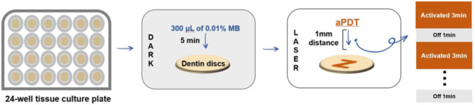
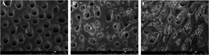
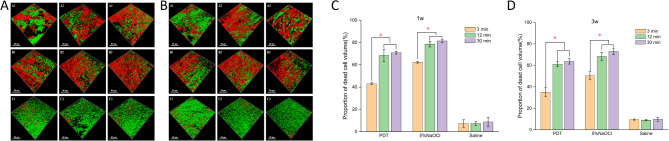
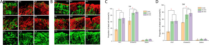
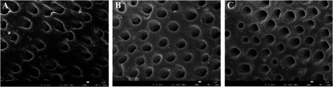
Methods: Prepared bovine and human dentin blocks and their structure were observed by scanning electron microscopy (SEM). Semicylindrical bovine dentin blocks and human root canal dentin blocks were inoculated with E. faecalis and incubated in air to form 1- and 3-week-old biofilms. The biofilms in dentin were subjected to aPDT with 0.01% MB, 5% NaOCl and saline with the exposure of 3, 12 and 30 min. The dead portions of bacterial cells in E. faecalis biofilms were analyzed with using LIVE/DEAD bacteria viability staining and CLSM.
Results: A clean dentin surface in bovine dentin blocks were verified with SEM. In bovine and human dentin blocks, significantly more bacteria were dead when aPDT with MB and 5% NaOCl were used with the long exposure time (12 and 30 min) than with 3 min (P < 0.05). The speed of killing was fastest during the first 3 min, and few more bacterial cells were killed after 12 min in the disinfection groups. 5% NaOCl exhibited the highest effectiveness of bacterial killing in dentin at each time point than aPDT with MB groups (P < 0.05). The proportion of killed bacteria was higher in young biofilms than in mature biofilms in aPDT with MB and NaOCl groups (P < 0.05). Moreover, there were no clearly visible changes in structure of bovine dentin surfaces subjected to aPDT with MB for 30 min.
Conclusion: aPDT with 0.01% MB has the capability to kill bacterial cells in E. faecalis biofilms on bovine and human dentin blocks. Young E. faecalis biofilms in dentin canals were more susceptible to disinfection approaches than mature biofilms.
Keywords: Enterococcus faecalis; Bacterial biofilms; Confocal laser scanning microscopy; Photodynamic therapy; Root canal disinfection.
© 2025. The Author(s).
Conflict of interest statement
Declarations. Ethics approval and consent to participate: Ethical approval The study was conducted according to the guidelines of the Declaration of Helsinki, and approved by the Science and Ethics Commission of the First Affiliated Hospital of Zhengzhou University. Consent for publication: Not applicable. Competing interests: The authors declare no competing interests. Informed consent: Not applicable.
Figures
References
-
- Fulghum RS, Wiggins CB, Mullaney TP. Pilot study for detecting obligate anaerobic bacteria in necrotic dental pulps. J Dent Res. 1973;52:637. - DOI
MeSH terms
Substances
Grants and funding
LinkOut - more resources
Full Text Sources
Medical
Miscellaneous
