. 2025 Feb 22;11(1):34.
doi: 10.1038/s41522-025-00645-6.

Hypertension inhibition by Dubosiella newyorkensis via reducing pentosidine synthesis

Affiliations

Hypertension inhibition by Dubosiella newyorkensis via reducing pentosidine synthesis

Tian-Hao Liu et al. NPJ Biofilms Microbiomes. .

Abstract

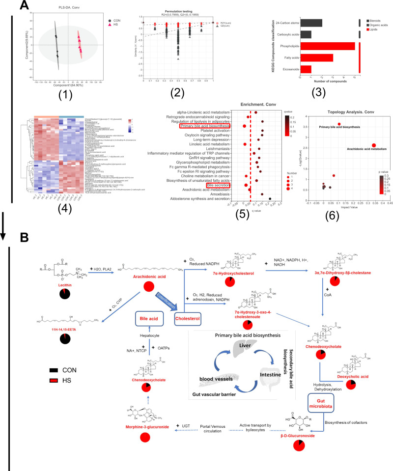
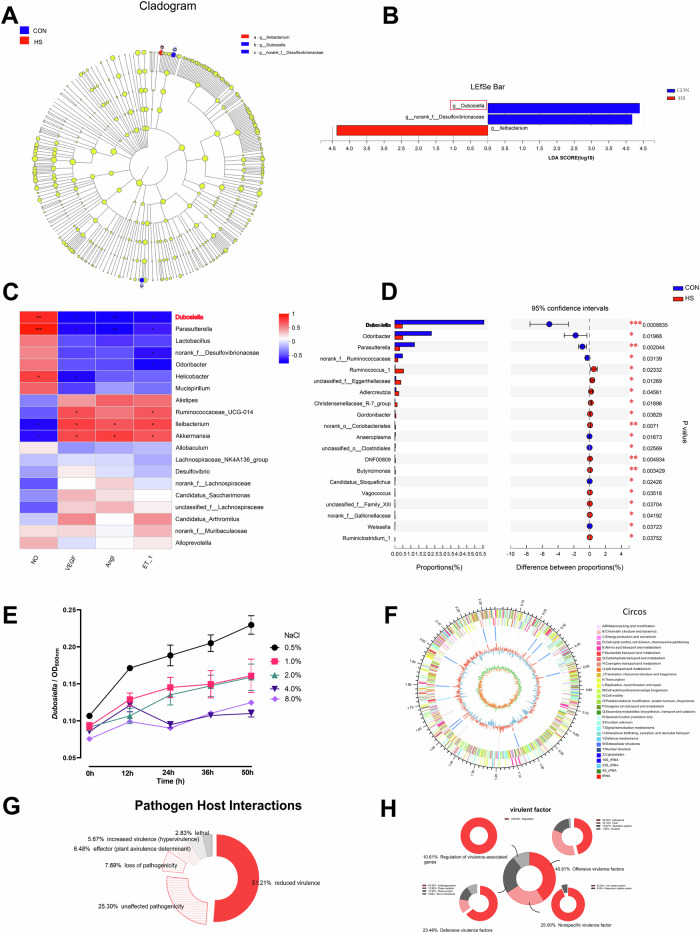
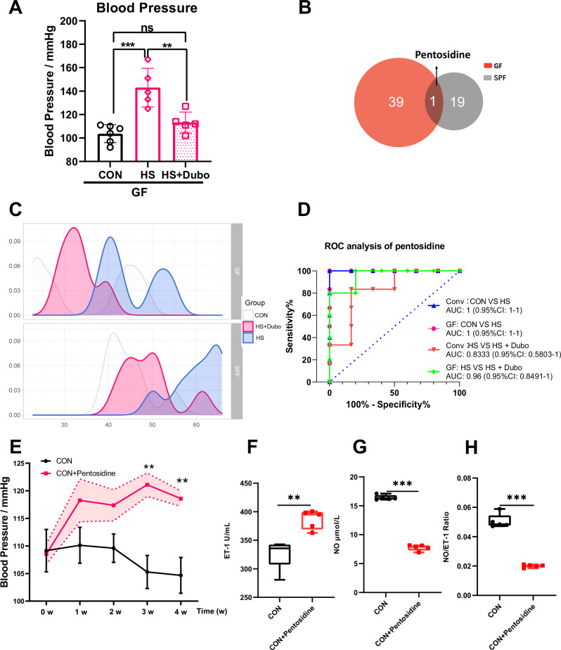
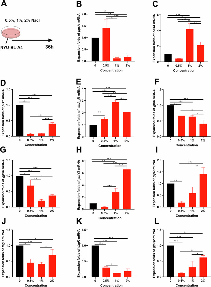
Gut dysbiosis has been associated with hypertension. Herein, we aimed to discover the potential association between gut microbiota and high-salt diet (HSD) induced endothelial dysfunction in conventional hypertensive mice. Dubosiella newyorkensis was found highly sensitive to salt in HSD-induced hypertension. The salt-sensitive nature of Dubosiella newyorkensis was confirmed by bacteria culture in vitro. Oral Dubosiella newyorkensis in HSD-induced hypertensive mice decreased blood pressure, inhibited activation of vascular endothelium, attenuated inflammation and alleviated intestinal vascular barrier injury. Similar effects of Dubosiella newyorkensis were observed in germ-free mice. Interestingly, serum pentosidine was found to function as a biomarker for Dubosiella newyorkensis in response to HSD in both metabolic modes. Supplement of pentosidine, deteriorated hypertension and vascular endothelial damage. Differential genes enriched in the glycerophospholipid metabolism were markedly altered in cultured bacteria. Our study has identified Dubosiella newyorkensis as a new salt-sensitive gut microbe that inhibits pentosidine production thereby alleviating hypertension.


Conflict of interest statement

Competing interests: The authors declare no competing interests.

Figures

Fig. 1
Fig. 1. Ratio of plasma NO/ET-1 decreased hypertension by high-salt diet (HSD) in mice.
A The changes in blood pressure (n = 10). BD The changes in vasoconstriction and vasodilation-related factors, such as NO (B), ET-1 (C), and NO/ET-1 (D). EG The changes in vascular endothelial active factors, such as EMPs (E), VEGF (F), and Ang1 (G). HJ The changes in immunity and inflammation-related factors, such as TNF-α (H), IL-1β (I), IL-6 (J), and sIgA (K). LN The changes in the intestinal vascular barrier, such as NHE3 (L), DAO (M), and intestinal perfusion (N). The intestinal perfusion in the four groups (n = 3) was evaluated with laser speckle flowmetry, with red indicating high perfusion and high flux and blue indicating low perfusion and low flux. CON control group, HS high-salt diet group, NO nitric oxide, ET-1 endothelin-1, EMPs endothelial microparticles, VEGF vascular endothelial growth factor, AngI angiotensin I, IL-1β interleukin-1β, IL-6 interleukin-6, TNF-α tumor necrosis factor-α, sIgA secretory immunoglobulin A, DAO diamine oxidase, NHE3 Na+/H+ exchanger type 3. Data are presented as mean ± SEM. Statistical significance was determined using t-test. *p < 0.05, **p < 0.01, and ***p < 0.001; ns, no statistical significance; n = 6–7.
Fig. 2
Fig. 2. Metabolic profiles changed in hypertension.
A Metabolite differences in conventional (Conv) mice induced by HSD (n = 6). A1 The PLS-DA plot. A2 Permutation testing of the PLS-DA analysis. A3 The KEGG compound classification. A4 The heatmap of metabolites. A5 The enrichment of metabolites. A6 The topology analysis. B The metabolic cycle pathway of the HSD in Conv mice based on KEGG mapping of Genome. Net: map00120 with a 4/21 ratio of metabolites annotated to the pathway, including chenodeoxycholate (C02528), 7alpha-hydroxycholesterol (C03594), 3alpha,7alpha-dihydroxy-5beta-cholestane (C05452), and 7alpha-hydroxy-3-oxo-4-cholestenoate (C17337) (q < 0.05); map0497 with a 4/21 ratio of metabolites annotated to the pathway, including beta-D-glucuronoside (C03033), deoxycholic acid (C04483), morphine-3-glucuronide (C16643), and chenodeoxycholate (C02528) (q < 0.05); map00590 with a 3/21 ratio of metabolites annotated to the pathway, including arachidonate (C00219), lecithin (C00157), and 11H-14,15-EETA (C14813) (q < 0.05). CON control group, HS high-salt diet group.
Fig. 3
Fig. 3. Potential probiotic Dubosiella is highly sensitive to salt in hypertension.
A, B LDA effect size (LEfSe) analysis between subsets of high-salt diet in conventional (Conv) mice, LDA = 4. C The correlation analysis of bacteria genus and vascular endothelial factors. D The changes in the relative abundance of Dubosiella after 4 weeks of high-salt diet treatment in Conv mice. E The salt tolerance test results of Dubosiella newyorkensis. F The whole Dubosiella newyorkensis genome. From the outer to the inner circle, each circle indicates information regarding the genome of the forward CDS, reverse CDS, forward COG function classification, reverse COG function classification, nomenclature and locations of predictive secondary metabolite clusters, and G+C content. The red sections indicate that the GC content of the region is higher than the average GC content of the whole genome. The higher the peak value, the greater the difference between the GC content and the average GC content. The blue sections indicate that the GC content of the region is lower than the average GC content of the whole genome. The higher the peak value, the greater the difference between the GC content and the average GC content and GC skew. G The analysis of pathogen–host interactions. H The analysis of virulence genes. Statistical significance was determined using t-test. *p < 0.05, **p < 0.01, and ***p < 0.001; n = 6.
Fig. 4
Fig. 4. Supplementing Dubosiella newyorkensis prevents the rise of blood pressure in hypertensive mice.
A The changes in blood pressure (n = 10). BD The changes in vasoconstriction and vasodilation-related factors, such as NO (B), ET-1 (C), and NO/ET-1 (D). EG The changes in vascular endothelial active factors, such as EMPs (E), VEGF (F), and Ang1 (G). HK The changes in immunity and inflammation-related factors, such as TNF-α (H), IL-1β (I), IL-6 (J), and sIgA (K). LN The changes in the intestinal vascular barrier, such as NHE3 (L), DAO (M), and intestinal perfusion (N). The intestinal perfusion in the four groups (n = 3) was evaluated with laser speckle flowmetry, with red indicating high perfusion and high flux and blue indicating low perfusion and low flux. HS high-salt diet group, HS+Dubo high-salt diet with supplementary NYU-BL-A4 group, NO nitric oxide, ET-1 endothelin-1, EMPs endothelial microparticles, VEGF vascular endothelial growth factor, AngI angiotensin I, IL-1β interleukin-1β, IL-6 interleukin-6, TNF-α tumor necrosis factor-α, sIgA secretory immunoglobulin A, DAO diamine oxidase, NHE3 Na+/H+ exchanger type 3. Data are presented as mean ± SEM. Statistical significance was determined using t-test. *p < 0.05, **p < 0.01, and ***p < 0.001; ns, no statistical significance; n = 6–7.
Fig. 5
Fig. 5. Pentosidine, production inhibited by Dubosiella newyorkensis, increases blood pressure in mice.
A The changes in blood pressure of GF mice. B, C The common serum metabolite was screened using the Venn diagram and density curve. D ROC analysis of pentosidine in HSD-induced Conv and GF mice with supplementary NYU-BL-A4. E The changes in blood pressure with supplementary pentosidine. F, G, H The changes in vasoconstriction and vasodilation-related factors, such as NO, ET-1, and NO/ET-1. CON control group, HS high-salt diet group, HS+Dubo high-salt diet with supplementary NYU-BL-A4 group, CON+Pentosidine supplementary pentosidine group. Data are presented as mean ± SEM. One-way ANOVA was used for multi-group comparison, and t-test was used for the two groups. *p < 0.05, **p < 0.01, and ***p < 0.001; ns, no statistical significance; n = 5–6.
Fig. 6
Fig. 6. Dubosiella newyorkensis decreases blood pressure by modulating the synthesis of pentosidine in mice.
A The PCA plot. B The heatmap of metabolites in Conv and GF mice. C1 The Venn diagram in Conv mice. C2 The heatmap of differential metabolites in Conv mice. D1 The Venn diagram in GF mice. D2 The heatmap of differential metabolites in GF mice. E Dubosiella newyorkensis (NYU-BL-A4) altered the common serum metabolic pathway in conventional (Conv) and germ-free (GF) mice fed a high-salt diet, and in the shared pathways, the upregulated pathways are marked in green and the downregulated pathways in red. HS high-salt diet, Dubo/DUBO, Dubosiella newyorkensis (NYU-BL-A4).
Fig. 7
Fig. 7. Differential genes enriched in the screened glycerophospholipid metabolism pathway were observed in vitro bacterial experiment.
A Design of bacterial experiments. BL The relative mRNA levels of the 11 differential genes in the glycerophospholipid metabolism pathway. Data are presented as mean ± SEM. Statistical significance was determined using one-way ANOVA. *p < 0.05, **p < 0.01, and ***p < 0.001; n = 3.
Fig. 8
Fig. 8. Schematic diagram depicting the response of gut microbiota to high-salt diet (HSD) in mice by modulating the synthesis of pentosidine.
① HSD-induced increased blood pressure in conventional (Conv) mice and the gut symbiotic bacteria Dubosiella newyorkensis (NYU-BL-A4) was screened; ② Dubosiella newyorkensis (NYU-BL-A4) played an important role in HSD-induced mice; ③ The potential mechanism of action has been explored; ④ The primary metabolic route and important metabolites have been verified in vivo animal research and in vitro bacterial experiments. Dubo Dubosiella newyorkensis (NYU-BL-A4), NO nitric oxide, ET-1 endothelin-1, EMPs endothelial microparticles, VEGF vascular endothelial growth factor, AngI angiotensin I, IL-1β interleukin-1β, IL-6 interleukin-6, TNF-α tumor necrosis factor-α, sIgA secretory immunoglobulin A, DAO diamine oxidase, NHE3 Na+/H+ exchanger type 3, Conv conventional, GF germ-free.

References

    1. He, F. J., Campbell, N., Woodward, M. & MacGregor, G. A. Salt reduction to prevent hypertension: the reasons of the controversy. Eur. Heart J.42, 2501–2505 (2021). - PubMed
    1. O’Donnell, M. et al. Salt and cardiovascular disease: insufficient evidence to recommend low sodium intake. Eur. Heart J.41, 3363–3373 (2020). - PubMed
    1. Chen, L. et al. Modest sodium reduction increases circulating short-chain fatty acids in untreated hypertensives a randomized, double-blind, placebo-controlled trial. Hypertension76, 73–79 (2020). - PMC - PubMed
    1. Kovacic, J. C. et al. Endothelial to mesenchymal transition in cardiovascular disease: JACC state-of-the-art review. J. Am. Coll. Cardiol.73, 190–209 (2019). - PMC - PubMed
    1. Wilck, N. et al. Salt-responsive gut commensal modulates T(H)17 axis and disease. Nature551, 585 (2017). - PMC - PubMed

LinkOut - more resources