. 2025 Feb 22;8(1):278.
doi: 10.1038/s42003-025-07662-3.

Distinct outcomes from targeted perturbations of the multi-subunit SCFSkp2 E3 ubiquitin ligase in blocking Trp53/Rb1-null prostate tumorigenesis

Affiliations

Distinct outcomes from targeted perturbations of the multi-subunit SCFSkp2 E3 ubiquitin ligase in blocking Trp53/Rb1-null prostate tumorigenesis

Yingjiao Xue et al. Commun Biol. .

Abstract

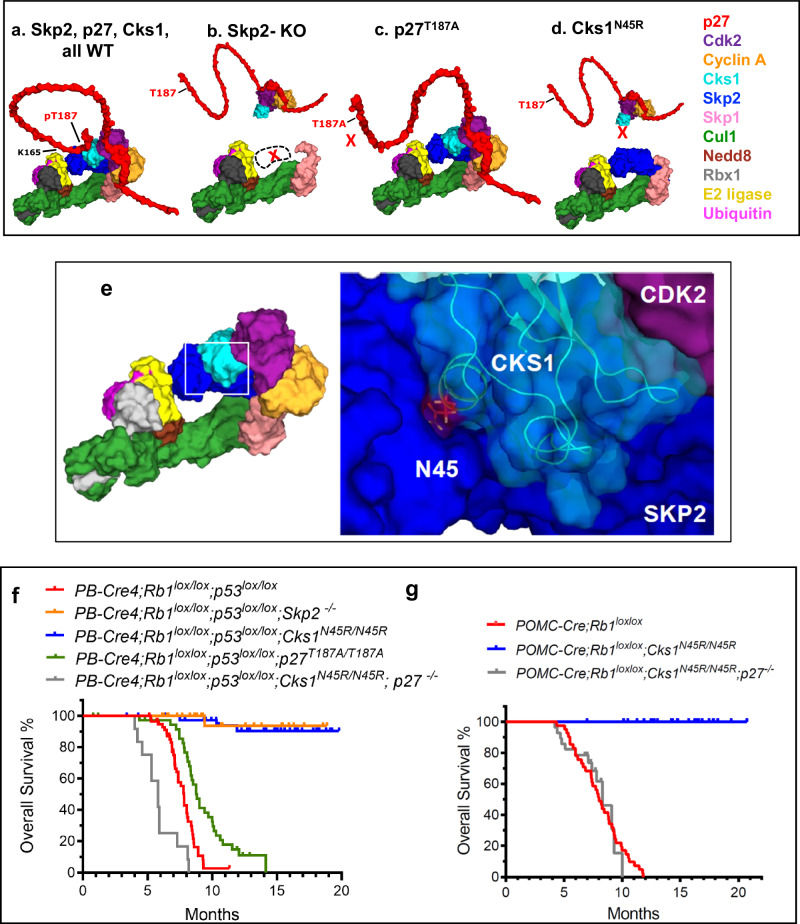
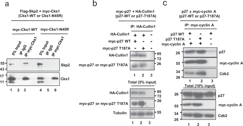
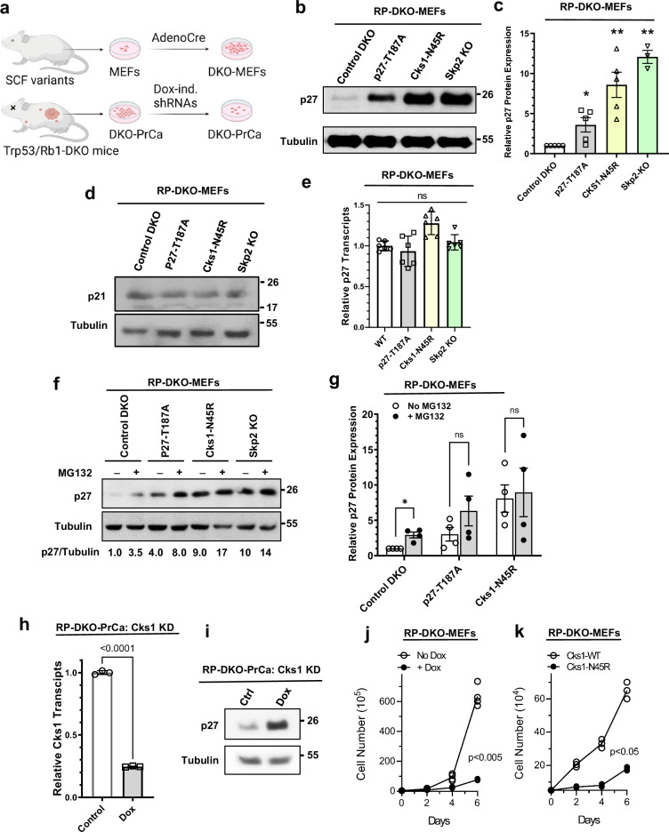
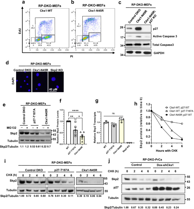
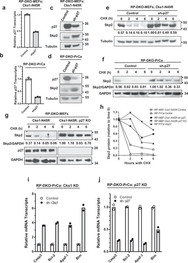
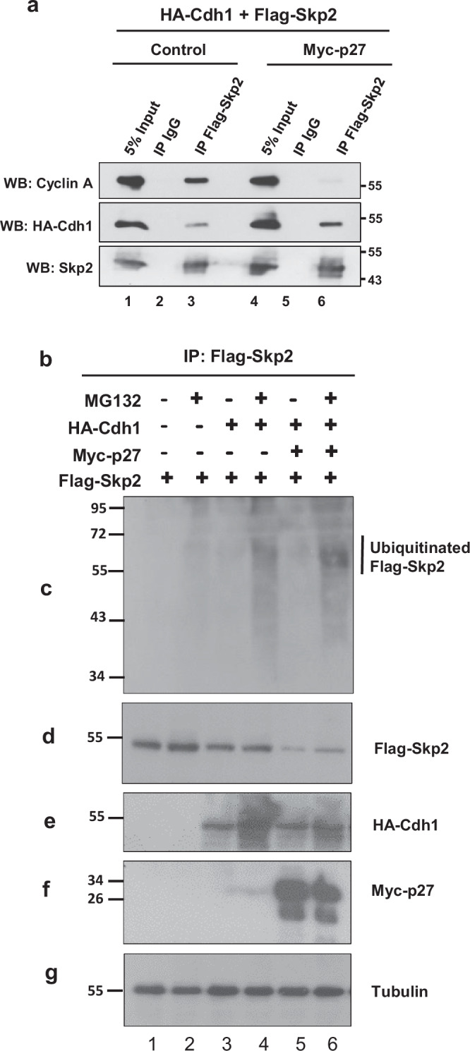
Identifying effective therapies targeting multi-protein complexes that lack catalytic sites or cofactor pockets remains a long-standing challenge. The proto-oncogene, ubiquitin E3 ligase SCFSkp2, is one such target. SCFSkp2 promotes the proteasomal degradation of the cyclin-dependent kinase inhibitor p27, which controls cell cycle progression. Targeted knockout of Rb1/Trp53 causes metastatic prostate cancer in mice; additional knockout of Skp2 completely blocks tumorigenesis. We compared gene-edited mice that carried two different single amino acid changes in the SCFSkp2 complex, structurally predicted to inhibit the degradation of p27. Mutation of the SCFSkp2 accessory protein Cks1 (Cks1N45R) completely blocked Rb1/Trp53-driven prostate tumorigenesis, phenocopying Skp2 knockout, whereas a mutation directly stabilizing p27 (p27T187A) did not. This was consistent with structural models that predicted the binding of both p27 and p27T187A to the SCFSkp2/Cks1/Cdk2/CyclinA/p27 complex, and their subsequent ubiquitination and degradation, albeit at different rates. Two binding modes, which differ in their dependence on phosphorylated T187, are predicted by the model. Studies confirmed the role of p27 in mediating tumorigenesis in Rb1/Trp53 mutant tumors and revealed a mutually destabilizing Skp2 and p27 feedback loop. The integration of gene editing, drug-surrogate mutations, and mouse tumor models offers a blueprint for studying SCFSkp2 and other multi-subunit biomedical targets.


Conflict of interest statement

Competing interests: The authors declare the following competing interests: E.S. is a consultant for Sandoz Pharmaceuticals. J.P.M. is a co-founder of Exo Therapeutics, a company focused on substrate-binding sites. The remaining authors declare no competing interests.

Figures

Fig. 1
Fig. 1. Stepwise binding and post-translational events involved in the ubiquitin-dependent proteasomal degradation of p27 mediated by SCFSkp2.
A SCFSkp2 has a strong binding affinity for Cks1 to form SCFSkp2-Cks1 in early S-phase when CDK2-Cyclin complexes are predominantly bound to the N-terminus of p27 cyclin-binding motif,. The intrinsic disorder of the N- and C-termini of p27 are highlighted with a circular arrow over the crystallographically unresolved residues 1–26 and 94–198,. B The CDK2-Cyclin-p27 complexes require Cks1 to associate with SCFSkp2-Cks1 (or may together recruit free p27 after step G, not shown). C Y88/Y89 and Y74 are tyrosine residues known to require phosphorylation by non-receptor tyrosine kinases to enable the maximum rate of T187 phosphorylation by a proximal CDK2 enzyme bound to Cks1 or to the N-terminus of Skp2. D The phosphorylation of pT187 provides a critical negative charge enabling the C-terminus of p27 to associate with the phosphodegron site of SCFSkp2-Cks1, which is a binding groove comprised of Skp2 and Cks1 residues. E Binding of the p27(pT187) phosphodegron (residues 181–190) brings the intrinsically disordered residues of p27 in proximity to the E2 enzyme to facilitate the first ubiquitin transfer; for example, as shown here on K165, among other p27 Lysine residues. F Subsequent ubiquitin-K48-ubiquitination (Ub’ and Ub”) events may occur independently of the phosphodegron site, but the p27 C-terminal region is likely to be sufficiently flexible to associate with the phosphodegron site in the presence of oligo-ubiquitin chains as shown by this model. G Finally, poly-ubiquitinated p27 is recruited and loaded into the proteasome for proteolytic degradation (shown in a cartoon). The posttranslational change occurring in each step is highlighted with boldface.
Fig. 2
Fig. 2. Distinct outcomes from targeted genetic perturbations of the multi-subunit SCFSkp2 E3 ligase in Rb1-loss driven tumorigenesis models.
ad Three-dimensional models of the higher order complexes of SCFSkp2/Cks1/Cdk2/cyclinA/p27 having all wildtype proteins (a); and representations of a complex that cannot form due to lack of Skp2 (b); of a complex binding the p27T187A variant (c); and of the effect of the Cks1N45R mutation on complex formation (d). The intrinsically disordered sections of p27 are represented as loops (Fig. 1). e Interface of Skp2-Cks1 highlighting the pocket occupied by the residue N45 that is changed in the gene-edited subunit Cks1(N45R),. f, g Kaplan-Meier survival curves in mice with Rb1/Trp53 double knock out-driven prostate cancers (f), and with Rb1-knockout driven pituitary cancers (g), when combined with the respectively indicated genotypes and targeted perturbations. Includes some data for Skp2 KO and p27T187A mice that have been previously published by our group. The size of each cohort is noted in Table 1. f All groups were significantly different from Rb1/Trp53-DKO, p < 0.0001 by Log-rank test. g POMC-Cre;Rb1lox/lox was significantly different POMC-Cre;Rb1lox/lox; Cks1N45R/N45R.
Fig. 3
Fig. 3. Comparison of the binding of Cks1N45R and p27T187A to Skp2, Cyclin A, and Cdk2.
a Immunoprecipitation of Myc-Cks1 from 293 T cells overexpressing Flag-Skp2 and Myc-Cks1-wild type or mutant Myc-Cks1N45R. Western blot shows loss of coimmunoprecipitated Skp2 in cells expressing Cks1N45R (compare lanes 6–3). b 293 T cells were transfected with HA-Cullin1 and either wildtype p27 or p27T187A. IP was with HA-Cullin1 followed by Western blot with HA-Cullin1 and myc-p27. c 293 T cells were transfected with either wildtype p27 or p27T187A, with or without Myc-cyclin A. IP was with Myc-cyclin A, followed by Western blot for p27, Myc-Cyclin A or Cdk2.
Fig. 4
Fig. 4. Cks1N45R knock-in is significantly more effective than p27T187A knock-in in blocking p27 proteasomal degradation.
a Derivation of Rb1/Trp53 double-knockout mouse embryonic fibroblast cells (RP-DKO-MEFs) used in this study. Image was created in BioRender (Maianti J., 2025 https://BioRender.com/n34z903). b Western blot of p27 in RP-DKO-MEFs of four genotypes, as indicated. c Quantitation of p27 protein levels as shown in (b) (n = five biologically independent western blots). * Indicates significantly different from control, ** significantly different than control and p27-T187A. Mean ± SEM, n = 3–5. d Western blot of the CIP/KIP protein p21 in RP-DKO-MEFs. e p27 mRNA levels (relative to GAPDH) of RP-DKO-MEFs corresponding to western in (d). ns, not significant. Mean ± SEM, n = 6 biologically independent samples. f p27 levels in RP-DKO-MEFs of four genotypes treated with or without MG132 (20 μM) for 6 hours. g Quantitation of levels of p27 protein (corrected for tubulin) as shown in (f). * Indicates significant effect of MG132. Mean ± SEM, n = 4 biologically independent samples. h, i Early passage primary prostate tumor cells from Rb1 and Trp53 double knockout mice (RP-DKO-PrCa cells) were transfected with a pTripZ-shCks1 construct. Cks1 knockdown was induced by doxycycline. h Cks1 mRNA levels were measured by RT-qPCR. Mean ± SEM, n = 3 biologically independent samples. i Western blot showing an increase in p27 protein level upon Cks1 knockdown. j Cell proliferation in vitro of RP-DKO-PrCa cells with or without doxycycline treatment to knock down Cks1. Mean ± SEM, n = 3 biologically independent samples. k Cell growth in vitro of RP-DKO-MEFs with wild type Cks1 or Cks1N45R. Mean ± SEM, n = 3 biologically independent samples.
Fig. 5
Fig. 5. Cks1N45R induces apoptosis and causes cell cycle arrest in G1 and S phases, in a p27-dependent manner.
a, b Cell cycle distribution and quantitation of Cks1-WT (a) and Cks1N45R (b) in RP-DKO-MEFs as analyzed by flow cytometry (PI, propidium iodide; EdU, 5-ethynyl-2´-deoxyuridine). Distribution of cells in sub-G1, G1, S, and G2-M are indicated. c Western blot of active caspase 3 levels in RP-DKO MEFs with Cks1-WT, Cks1N45R, and combined Cks1N45R with p27 KO. d Immunofluorescence of nuclei of RP-DKO-MEFs stained with DAPI. e Western blot of Skp2 in RP-DKO-MEFs of indicated genotypes with and without MG132. f Quantification of Skp2 protein levels of RP-DKO-MEFs as shown in (e) (Mean ± SEM, n = 6 biologically independent western blots). g Corresponding Skp2 mRNA in RP-DKO-MEFs. ns, not significant. Mean ± SEM, n = 3 biologically independent samples. h, i Skp2 protein stability in RP-DKO-MEFs was determined and quantified by treating cells with cycloheximide (CHX) for the indicated times. j Skp2 and p27 protein in RP-DKO-PrCa cells with Cks1 knock down and cycloheximide.
Fig. 6
Fig. 6. Knockdown of p27 causes increased Skp2 protein due to a reduction in Skp2 proteasomal degradation.
a, b p27 was knocked down in RP-DKO-Cks1N45R MEFs (a) and RP-DKO-PrCa cells (b), and p27 mRNA levels determined by RT-qPCR. Mean ± SEM, n = 3 biologically independent samples. c, d Western blots of Skp2 and p27 from cells in (a, b). e, f Cells treated with cycloheximide (CHX) to determine the degradation rates of Skp2. Effect of p27 knockdown in Cks1N45R RP-DKO-MEFs (e) and RP-DKO-PrCa (f). g Skp2 protein levels in Cks1N45R RP-DKO-MEFs with and without p27 knockout, and with CHX. h Quantification of Skp2 degradation from (eg). i, j Effect of Cks1 knock down (i) and Skp2 knock down (j) in RP-DKO-PrCa cells on mRNA levels of the indicated E2F1-regulated apoptotic genes, relative to GAPDH mRNA. Mean of two biologically independent samples.
Fig. 7
Fig. 7. Increased p27 promotes Skp2 protein binding to APCCDH1 and APCCDH1-dependent Skp2 ubiquitination and proteasomal degradation.
a Coimmunoprecipitation of cyclin A or HA-Cdh1 with Flag-Skp2 in 293 T cells overexpressing Flag-Skp2, with (lanes 4–6) or without (lanes 1–3) Myc-p27 overexpression. bg 293 T cells were transfected with Flag-Skp2 (lanes 1–6), HA-Cdh1 (lanes 3–6), or Myc-p27 (lanes 5, 6), as indicated. All groups were treated with and without MG132. After IP of Skp2 with an anti-mouse Flag antibody, its ubiquitinated forms were detected with an anti-rabbit ubiquitin antibody (c). Ubiquitinated Flag-tagged Skp2 has an estimated molecular weight of 58-63 kDa. Total cell extracts were probed with an anti-mouse Flag (d), anti-rabbit HA (e), anti-mouse Myc (f), or anti-mouse tubulin antibodies (g).

Similar articles

References

    1. Armenia, J. et al. The long tail of oncogenic drivers in prostate cancer. Nat. Genet.50, 645–651 (2018). - PMC - PubMed
    1. Nava Rodrigues, D. et al. RB1 heterogeneity in advanced metastatic castration-resistant prostate cancer. Clin. Cancer Res.25, 687–697 (2019). - PubMed
    1. Mateo, J. et al. Genomics of lethal prostate cancer at diagnosis and castration resistance. J. Clin. Invest.130, 1743–1751 (2020). - PMC - PubMed
    1. Zhou, Z. et al. Synergy of p53 and Rb deficiency in a conditional mouse model for metastatic prostate cancer. Cancer Res.66, 7889–7898 (2006). - PubMed
    1. Zhao, H. et al. Skp2 deletion unmasks a p27 safeguard that blocks tumorigenesis in the absence of pRb and p53 tumor suppressors. Cancer Cell24, 645–659 (2013). - PMC - PubMed

MeSH terms