. 2025 Apr;39(4):929-941.
doi: 10.1038/s41375-025-02542-5. Epub 2025 Feb 22.

STING mediates increased self-renewal and lineage skewing in DNMT3A-mutated hematopoietic stem/progenitor cells

Affiliations

STING mediates increased self-renewal and lineage skewing in DNMT3A-mutated hematopoietic stem/progenitor cells

Jingru Huang et al. Leukemia. 2025 Apr.

Abstract

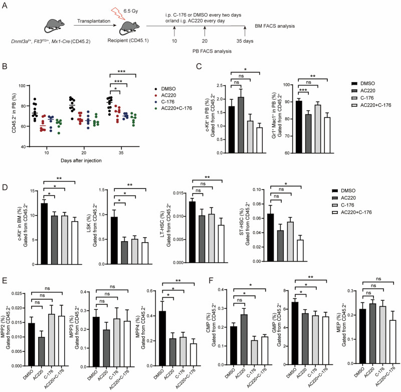
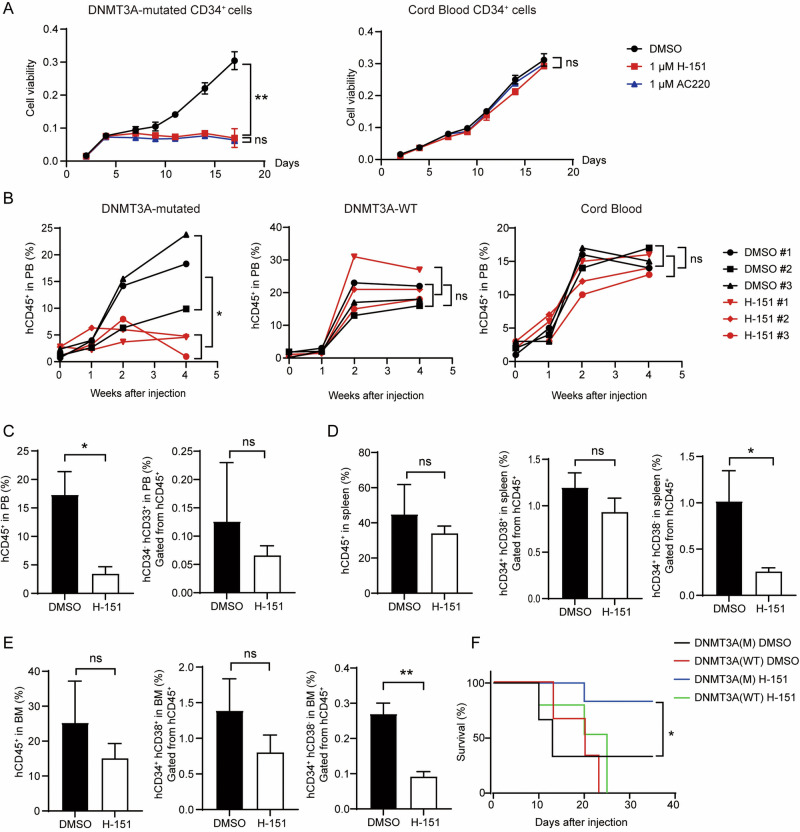
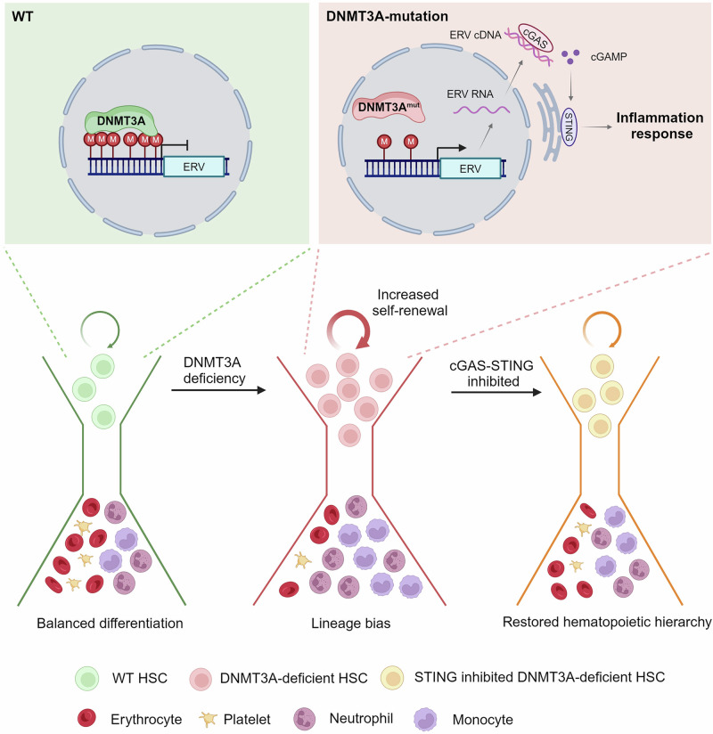
Somatic mutations in DNA methyltransferase 3 A (DNMT3A) are frequently observed in patients with hematological malignancies. Hematopoietic stem/progenitor cells (HSPCs) with mutated DNMT3A demonstrate increased self-renewal activity and skewed lineage differentiation. However, the molecular mechanisms underlying these changes remain largely unexplored. In this study, we show that Dnmt3a loss leads to the upregulation of endogenous retroviruses (ERVs) in HSPCs, subsequently activating the cGAS-STING pathway and triggering inflammatory responses in these cells. Both genetic and pharmacological inhibition of STING effectively corrects the increased self-renewal activity and differentiation skewing induced by Dnmt3a deficiency in mice. Notably, targeting STING showed inhibited acute myeloid leukemia (AML) development in a Dnmt3a-KO; Flt3-ITD AML model, comparable to AC220, an FDA-approved FLT3-ITD inhibitor. A patient-derived xenograft (PDX) model further demonstrated that targeting STING effectively alleviates the leukemic burden of DNMT3A-mutant AML. Collectively, our findings highlight a critical role for STING in hematopoietic disorders induced by DNMT3A mutations and propose STING as a potential therapeutic target for preventing the progression of DNMT3A mutation-associated leukemia.


Conflict of interest statement

Competing interests: The authors declare no competing interests. Ethics approval: All animal experiments described in this study were conducted in accordance with the ethical guidelines of the Department of Laboratory Animal Science Fudan University, No. 202307021S. The primary leukemia cells were collected from patient bone marrow cells and followed the ethical guidelines of Huashan Hospital, No. KY2015-269.

Figures

Fig. 1
Fig. 1. Loss of Sting impairs the increased self-renewal of HSPCs mediated by Dnmt3a deficiency.
A Schematic of the strategy used to generate Dnmt3a; Sting-DKO mice. The phenotypes of these mice were analyzed 16 weeks after poly(I:C) injection. B Deletion of Sting reduced the repopulating capacity of Dnmt3a-deficient LSK cells in CFU assays. LSK cells from different mice were sorted by FACS, and the number of CFUs was counted 7 days after plating. IFNβ was used at a concentration of 1 U/ml. C Loss of Sting suppressed the increased self-renewal of Dnmt3a-deficient LSK cells in vivo (n = 5). D The proportions of myeloid and lymphoid cells in peripheral blood were measured by FACS (n = 5). E Deletion of Sting specifically reduced the proliferation of Dnmt3a-KO HSPCs. The HSPCs were stained with an antibody against Ki67 and analyzed by FACS (n = 6). Data are presented as mean ± s.e.m., with *P < 0.05, **P < 0.01, ***P < 0.005, ****P < 0.0001, and “ns” not significant.
Fig. 2
Fig. 2. Deletion of Sting inhibits the development of CH associated with Dnmt3a deficiency in transplantation models.
A Diagram of the whole bone marrow transplantation assay. B Loss of Sting alleviated the splenomegaly in recipients of Dnmt3a-deficient donor bone marrow. The weight of spleens in recipient mice was measured 20 weeks after transplantation. Quantitative data are shown on the right (n = 8). C The plot graph shows the frequency of myeloid cells in the peripheral blood of recipient mice over the course of the transplantation assay (n = 11). D Deletion of Sting inhibited the myeloid differentiation bias mediated by Dnmt3a deficiency. The proportions of myeloid and lymphoid cells in peripheral blood were measured by FACS 20 weeks post-transplantation (n = 8). E Deletion of Sting restrained the self-renewal ability of Dnmt3a-deficient HSPCs. The proportions of LSK cells, LT-HSCs, ST-HSCs, CMPs, GMPs, and MEPs were measured by FACS. Gating plots are shown on the left, and quantitative data are shown on the right (n = 8). Data are presented as mean ± s.e.m., with *P < 0.05, **P < 0.01, ***P < 0.005, ****P < 0.0001, and “ns” not significant.
Fig. 3
Fig. 3. Inhibition of Sting reduces the self-renewal ability of Dnmt3a-deficient HSPCs.
A Diagram of competitive transplantation assay. B Injection of the STING inhibitor C-176 impaired the competitive advantage of Dnmt3a-deficient donors over the course of the competitive assay (n = 8). C Inhibition of STING alleviated the splenomegaly mediated by Dnmt3a deficiency (n = 5). D The bar graph shows the proportions of myeloid and lymphoid cells in the recipients (n = 5). E C-176 treatment reduced the proportion of Dnmt3a-deficient LSK cells in the recipients (n = 5). F The bar graph shows the frequencies of LT-HSCs, ST-HSCs, CMPs, GMPs, MEPs and MPP2/3/4 across different groups of recipient mice (n = 5). Data are presented as mean ± s.e.m., with *P < 0.05, **P < 0.01, ***P < 0.005 and “ns” not significant.
Fig. 4
Fig. 4. Dnmt3a deficiency induces the expression of ERVs in HSPCs.
A Pie charts display the percentages of upregulated repetitive elements in each class (top), and a heatmap shows the relative expression levels of the top 7 upregulated repetitive elements (bottom) across different Dnmt3a knockout HSPC lineages. B Deletion of Dnmt3a leads to hypomethylation of ERVs. The profile of average CpG methylation levels is displayed across regions including ERVs and their 2-kb flanking areas. The methylation levels of various ERV subfamilies are illustrated at the bottom. C The upregulation of ERVs in Dnmt3a-deficient c-Kit+ cells was measured using bulk RNA-seq (left) and the specific transcripts (right, top panel) and cDNA (right, bottom panel) of ERVs in c-Kit+ cells were quantified using q-PCR. A scatter plot illustrates the differentially expressed genes (circles) and transposable elements (triangles) between Dnmt3a-KOVav and WT c-Kit+ cells, with log2(fold change) >1 and P < 0.05 (n = 3). D Dnmt3a deficiency results in the hypomethylation of genic ERV loci in c-Kit+ cells. E FACS analysis showing the populations of specific cell types in the bone marrow of RTi-treated recipient mice (n = 5). Data are presented as mean ± s.e.m., with **P < 0.01, ***P < 0.005 and “ns” not significant.
Fig. 5
Fig. 5. Inhibition of STING restrains the leukemogenesis of mouse leukemia cells associated with Dnmt3a insufficiency.
A Diagram of comparing the biological activity of C-176 and FLT3 inhibitor AC220. B Injection of either C-176 or AC220 inhibited the myeloid differentiation bias of Dnmt3a-deficient leukemia cells over the course of the transplantation assay (n = 9). C Inhibition of STING restrained the expansion of naïve hematopoietic cells in the peripheral blood of recipient mice (n = 8). D C-176 and AC220 showed a combined effect in inhibiting the expansion of Dnmt3a+/−; Flt3ITD/+ leukemia stem cells (n = 8). E C-176 and AC220 specifically inhibited the expansion of MPP4 cells (n = 8). F C-176 and AC220 treatment reduced the proportions of CMP and GMP cells in Dnmt3a+/−; Flt3ITD/+ mice (n = 8). Data are presented as mean ± s.e.m., with *P < 0.05, **P < 0.01, ***P < 0.005 and “ns” not significant.
Fig. 6
Fig. 6. Targeting STING inhibits leukemia development in the PDX model.
A The effects of H-151 and AC220 treatments on patient-derived leukemia cells (left) and cord blood CD34+ cells (right) were evaluated in vitro using the CCK8 assay. B Inhibition of STING restrains the expansion of DNMT3A-mutated leukemia cells in NSG mice (n = 3 biological replicates). The DNMT3A-mutated patient sample harbors DNMT3A R882H and FLT3-ITD mutations, whereas the DNMT3A-WT sample carries CEBPA c.295_300del and TP53 P72R homozygous mutations. C Bar graphs display the proportions of human leukemia cells (hCD45+) and immature myeloid cells (hCD45+hCD34-hCD33+) (n = 3 biological replicates). Targeting STING diminishes the proportions of leukemia stem cells in the spleen (D) and bone marrow (E) of recipient mice. The proportions of human leukemia cells, blast cells (hCD45+hCD34+hCD38+), and leukemia stem cells (hCD45+hCD34+hCD38-) were analyzed via FACS (n = 3 biological replicates). F Survival analysis of recipient mice treated with DMSO or H-151, presented as a Kaplan–Meier curve (n = 3 with DMSO, 6 with H-151). Statistical significance was assessed by t-test (AE). Data are mean ± s.e.m., *P < 0.05; **P < 0.01.
Fig. 7
Fig. 7. Activation of the STING pathway by ERVs promotes increased self-renewal and skewed lineage populations in DNMT3A-mutated HSCs.
The mutated DNMT3A in HSCs leads to genome-wide hypomethylation. Beyond impacting gene regulation, this also results in the upregulation of previously silenced ERVs due to the absence of cytosine methylation-associated transcriptional inhibition. The increased ERV RNA can be reverse-transcribed into cDNA, which in turn activates the STING pathway and induces chronic inflammation in DNMT3A-mutated HSCs. This STING-dependent autonomous inflammation leads to increased self-renewal and biased lineage differentiation in DNMT3A-mutated HSCs. Inhibition of STING mitigates these phenotypes and prevents the development of leukemia associated with DNMT3A mutations in mouse models.

Similar articles

References

    1. Moore LD, Le T, Fan G. DNA Methylation and Its Basic Function. Neuropsychopharmacology. 2013;38:23–38. - PMC - PubMed
    1. Okano M, Bell DW, Haber DA, Li E. DNA Methyltransferases Dnmt3a and Dnmt3b Are Essential for De Novo Methylation and Mammalian Development. Cell. 1999;99:247–57. - PubMed
    1. Challen GA, Sun D, Jeong M, Luo M, Jelinek J, Berg JS, et al. Dnmt3a is essential for hematopoietic stem cell differentiation. Nat Genet. 2012;44:23–31. - PMC - PubMed
    1. Xie M, Lu C, Wang J, McLellan MD, Johnson KJ, Wendl MC, et al. Age-related mutations associated with clonal hematopoietic expansion and malignancies. Nat Med. 2014;20:1472–78. - PMC - PubMed
    1. Jaiswal S, Fontanillas P, Flannick J, Manning A, Grauman PV, Mar BG, et al. Age-related clonal hematopoiesis associated with adverse outcomes. N Engl J Med. 2014;371:2488–98. - PMC - PubMed