. 2025 Apr:354:199550.
doi: 10.1016/j.virusres.2025.199550. Epub 2025 Feb 21.

Adeno-associated virus 2 CRISPR/Cas9-mediated targeting of hepatitis B virus in tree shrews

Affiliations

Adeno-associated virus 2 CRISPR/Cas9-mediated targeting of hepatitis B virus in tree shrews

Md Haroon Or Rashid et al. Virus Res. 2025 Apr.

Abstract

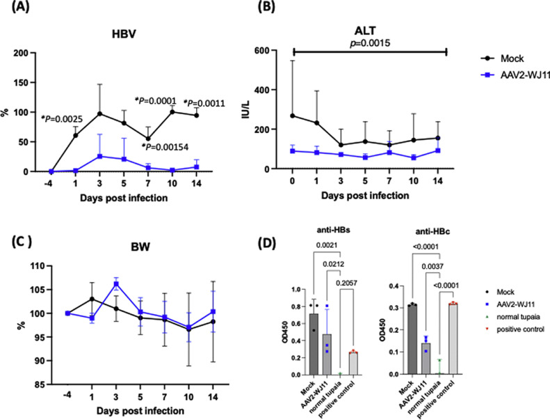
Chronic hepatitis B virus (HBV) infection is a global health issue with limited therapeutic options given the persistence of viral episomal DNA (cccDNA). Previously, we investigated the effects of adeno-associated virus 2 (AAV2) vector-mediated delivery of three guide (g)RNAs/Cas9 selected from 16 gRNAs. AAV2/WJ11-Cas9 effectively suppressed HBV replication in vitro and in humanized chimeric mouse livers. In the present study, we examined the effect of AAV2/WJ11-Cas9 on the acute phase of HBV genotype F infection in an immunocompetent northern tree shrew (Tupaia belangeri; hereafter, "tupaia") model. AAV2/WJ11-Cas9 treatment significantly reduced the HBV viral load in serum at 1, 7, 10, and 14 days post-infection (dpi). HBV-F infection caused enlargement of hepatocytes and mild lymphocytic infiltration in the interlobular connective tissue. Thus, the virus damages hepatocytes and drives infection progression and HBV core antigen (HBcAg) accumulation, which were not observed in AAV2/WJ11-Cas9 treated and normal liver tissues. AAV2/WJ11-Cas9 treatment reduced HBV DNA and cccDNA in liver tissues, as well as serum levels of HBV surface antigen and HBV core-related antigen (HBcrAg), including HBcAg and HBeAg at 14 dpi. Anti-HBc, anti-HBs, and anti-AAV Abs production was also detected. AAV2/WJ11-Cas9 treatment suppressed inflammatory cytokines and TLR1, TLR2, TLR3, TLR4, TLR6, TLR7, and TLR9 mRNA levels. Thus, WJ11/Cas9 delivered by AAV2 vectors may provide a new therapeutic approach for inhibiting HBV infection in immunocompetent animal models, which could be developed for use in humans through further translational research.

Keywords: Adeno-associated virus 2; HBV-F, CRISPR/Cas9; Hepatitis B virus; Northern tree shrew; Tupaia.


Conflict of interest statement

Declaration of competing interest The authors declare no conflict of interest.

Figures

Fig 1
Fig. 1
Experimental design of AAV2/WJ11-Cas9 and HBV genotype F mutant-type infection in adult tupaia. Adult tupaias (4–5 years) were either mock-treated or AAV2/WJ11-Cas9-treated, and HBV genotype F-Mt was simultaneously inoculated intravenously. Tupaia serum was collected −4, 1, 3, 5, 7, 10, and 14 days post infection (dpi).
Fig 2
Fig. 2
Effects of the AAV2/WJ11-Cas9 in the HBV-F-Mt-infected tupaia. (A) Detection of HBV DNA levels in serum at indicated time points. The level of HBV is indicated as a ratio to 10 dpi of HBV in mock-treated tupaia serum. A significant reduction in HBV levels was observed in AAV2/WJ11-Cas9-treated tupaia (n = 3) compared to that in non-treated controls (n = 3). (B) Serum ALT level, and (C) the ratio of body weight at indicated time points compared to day −4. (D) Anti-HBs and HBc antibodies were detected via ELISA in tupaia serum at 14 dpi with HBV infection. Error bars indicate the S.D. from three independent experiments. Statistical significance was calculated and p values <0.05 are indicated.
Fig 3
Fig. 3
Histopathological analysis of liver tissues obtained from mock-treated control (A: ×200 and B: ×400, rectangle in A), AAV2/WJ11-Cas9 treated (C:x200 and D:x400, rectangle in C), and uninfected control (E:x200 and F:x400, rectangle in E). (A) Approximately 50 % to 60 % of the hepatocytes in the liver tissue showed balloon-like swelling due to hydropic degeneration. (B) The cytoplasm of some swollen hepatocytes was slightly eosinophilic and exhibited a ground-glass appearance (blue arrow). Histologically, no significant changes were observed in the liver of the AAV2/WJ11-Cas9-treated tupaia (C and D) and normal tupaia (E and F). Detection of HBcAg in mock-treated (G) and AAV2/WJ11-treated (H) tupaia liver. Bars indicate 50 μm.
Fig 4
Fig. 4
HBV DNA (A) and HBV cccDNA (B) levels in the liver of mock-treated (n = 3) and AAV2/WJ11-Cas9-treated tupaia (n = 3) at 14 dpi. Serum HBsAg (C) and HBcrAg (D) levels in mock (n = 3) and AAV2/WJ11-Cas9-treated tupaia (n = 3) at 14 dpi. In both groups, the levels of Anti-HBs and Anti-HBc antibodies increased, indicating that the tupaias were immunocompetent (E) AAV genome expression in tupaia liver was measured via qPCR. (F) Immunoblot assay of tupaia liver tissues lyzed with a lysis buffer at a concentration of 10 % weight per volume was performed using 10 μL to detected Cas9 protein and actin (left). Protein bands were quantitated and normalized using the Fusion chemiluminescence imaging system (right). Statistical significance was calculated using the Student's t-test and p-values are indicated. Error bars indicate the S.D. from three independent experiments.
Fig 5
Fig. 5
Mutagenesis effect of AAV2/WJ11-Cas9 in tupaia liver. (A) PCR of HBV genome was performed using Q5 polymerase with HBV-Fmt819F and 2027R primers (Kayesh et al., 2020) (left, 1.2 kb fragment, blue arrowhead) from AAV2/WJ11-Cas9-treated tupaia liver genome (#329). T7E1 assay was conducted using purified and amplified HBV 1.2 kb fragment (right, blue, and orange arrowhead). A 1 kb DNA ladder marker (Nippon Genetics Co.) was used as a DNA molecular weight marker. (B) Sequence of targeting region of AAV2/WJ11-Cas9. The amplified HBV genome (1.2 kb) from genomic DNA of the tupaia liver was subcloned and sequenced (i-1). The WJ11 gRNA sequence is shown in red, and the PAMs are indicated in blue. The blue arrow indicates the predicted Cas9 cutting sites (For interpretation of the references to color in this figure legend, the reader is referred to the web version of this article.).
Fig 6
Fig. 6
TLR levels in liver tissues of mock-treated (n = 3) and AAV2/WJ11-Cas9-treated (n = 3) tupaia at 14 dpi. Statistical significance was calculated using the Student's t-test, and p values are indicated. Error bars indicate the S.D. from three independent experiments.

References

    1. Arauz-Ruiz P., Norder H., Robertson B.H., Magnius L.O. Genotype H: a new Amerindian genotype of hepatitis B virus revealed in Central America. J. Gen. Virol. 2002;83(Pt 8):2059–2073. - PubMed
    1. Bartosh U.I., Dome A.S., Zhukova N.V., Karitskaya P.E., Stepanov G.A. CRISPR/Cas9 as a new antiviral strategy for treating Hepatitis viral infections. Int. J. Mol. Sci. 2023;25(1) - PMC - PubMed
    1. Bello K.E., Mat Jusoh T.N.A., Irekeola A.A., Abu N., Mohd Amin N.A.Z., Mustaffa N., Shueb R.H. A recent prevalence of Hepatitis B virus (HBV) genotypes and subtypes in Asia: a systematic review and meta-analysis. Healthc. (Basel) 2023;11(7) - PMC - PubMed
    1. Cai B., Chang S., Tian Y., Zhen S. CRISPR/Cas9 for hepatitis B virus infection treatment. Immun. Inflamm. Dis. 2023;11(5):e866. - PMC - PubMed
    1. Cressman D.E., Greenbaum L.E., DeAngelis R.A., Ciliberto G., Furth E.E., Poli V., Taub R. Liver failure and defective hepatocyte regeneration in interleukin-6-deficient mice. Science. 1996;274(5291):1379–1383. - PubMed

Publication types

MeSH terms

Supplementary concepts

LinkOut - more resources