. 2025 Feb 8;53(4):gkaf102.
doi: 10.1093/nar/gkaf102.

Structural basis of endo-siRNA processing by Drosophila Dicer-2 and Loqs-PD

Affiliations

Structural basis of endo-siRNA processing by Drosophila Dicer-2 and Loqs-PD

Na Cao et al. Nucleic Acids Res. .

Abstract

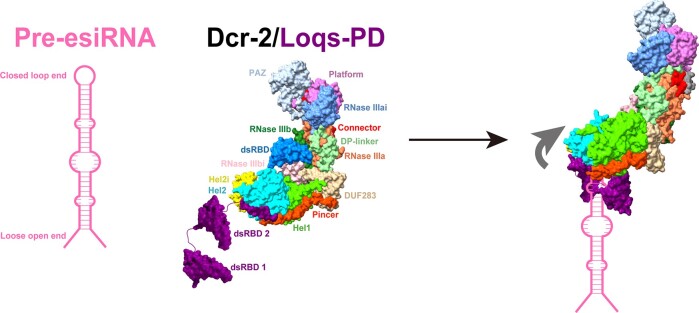
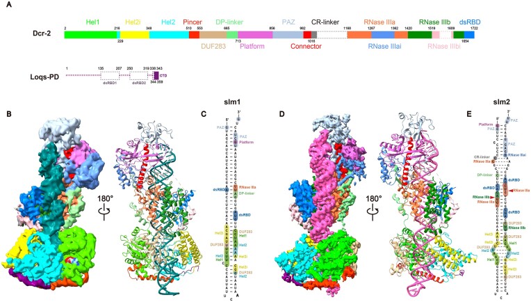
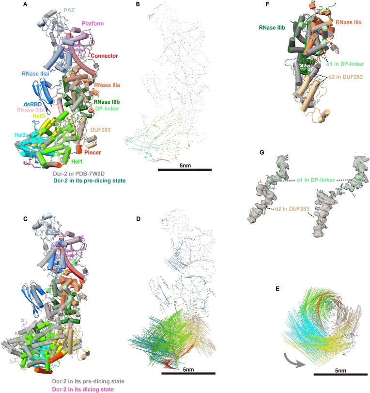
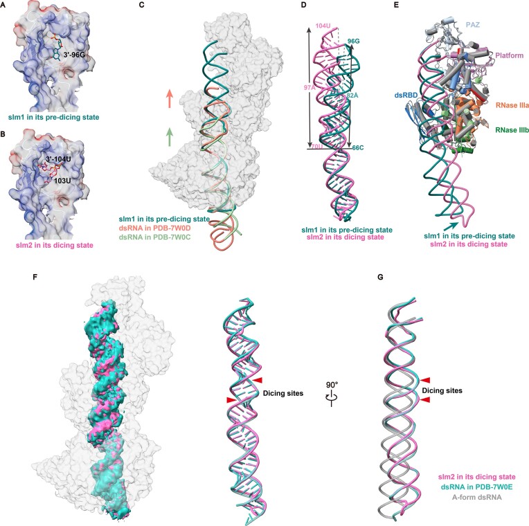
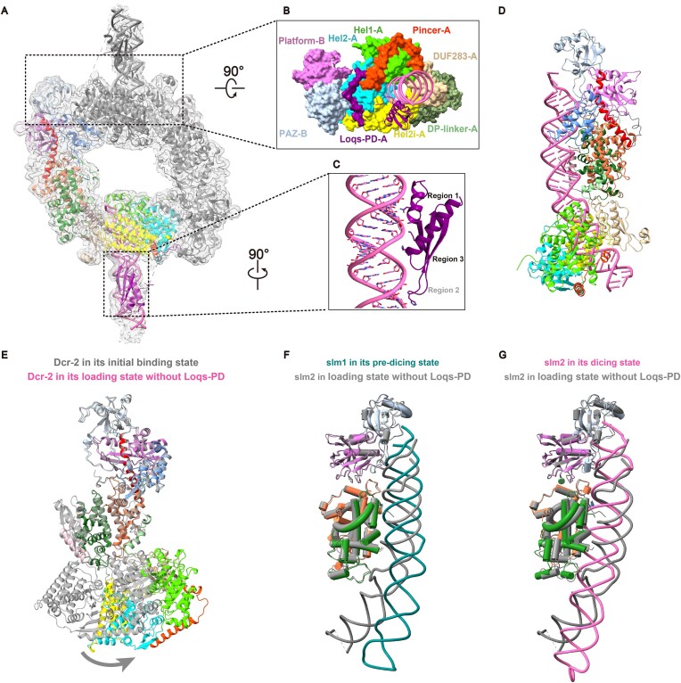
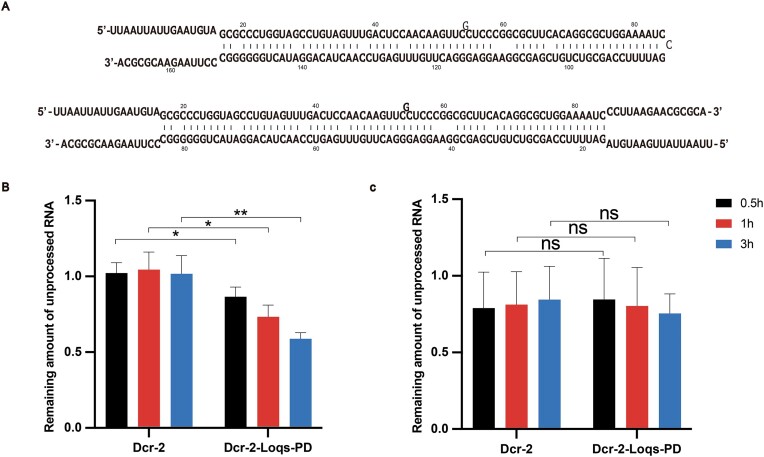
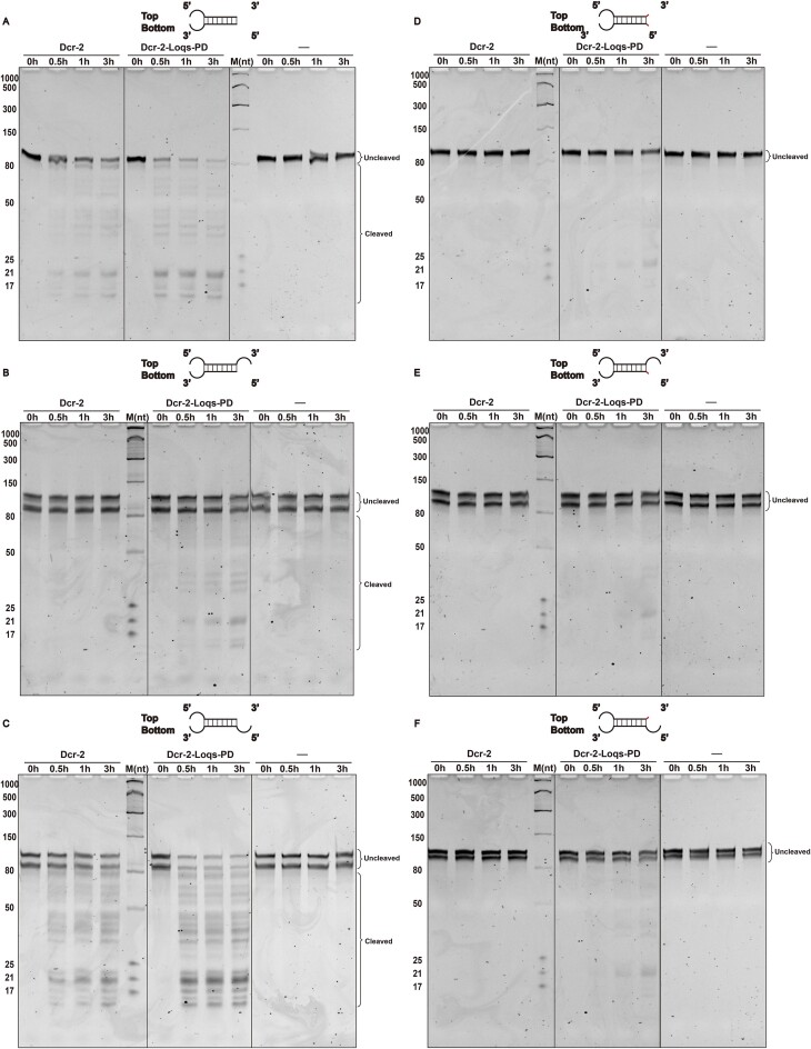
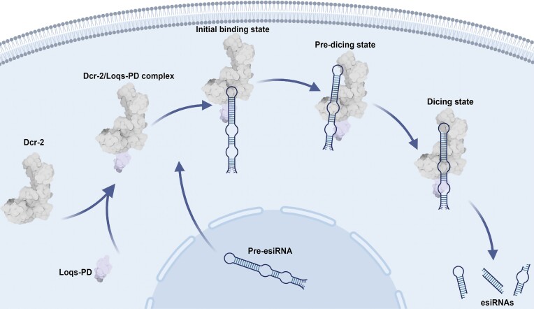
Endogenous small interfering RNAs (endo-siRNAs or esiRNAs) originate from either elongated endogenous transcripts capable of forming complex fold-back structures or from double-stranded regions generated through intermolecular base pairing of convergently transcribed mRNAs. The mechanism of maturation and functionality of esiRNAs exhibit significant variation across diverse species. In Drosophila melanogaster, esiRNAs reside in both somatic and germline cells, where they serve as post-transcriptional modulators for specific target RNAs. Their maturation process critically relies on Dicer-2 (Dcr-2), with the assistance of its cofactor Loqs-PD. In this study, we have successfully elucidated the cryo-EM structures of Dcr-2/Loqs-PD complex bound to esiRNA precursors (pre-esiRNAs) in various states. Our structural and biochemical results reveal that ATP is essential for the cleavage of esiRNAs by the Dcr-2/Loqs-PD complex, a process analogous to the cleavage of double-stranded RNA (dsRNA). When Loqs-PD is present, pre-esiRNAs are preferentially loaded onto the Helicase domain of Dcr-2. Moreover, as the Helicase domain exhibits a preference for binding to the rigid end of double-stranded RNA, Dcr-2 tends to cleave pre-esiRNA from the small closed loop end, rather than the loose and flexible open end.


Conflict of interest statement

None declared.

Figures

Graphical Abstract
Graphical Abstract
Figure 1.
Figure 1.
Cryo-EM structures of the Dcr-2/Loqs-PD complex in the dicing and pre-dicing states with pre-esiRNAs. (A) Schematic illustration of the domain arrangement of Dcr-2 and Loqs-PD. Disordered segments are represented by dashed lines. (B) Overall cryo-EM density map and cartoon model of Dcr-2/Loqs-PD/slm1 in the pre-dicing state. (C) Schematic diagram showing the interactions between the Dcr-2 and slm1 in the pre-dicing state. The bases involved in Watson–Crick base pairing are connected with a solid line, whereas the rest are placed separately or connected through dashed lines [same for (E)]. (D) Overall cryo-EM density map and cartoon model of Dcr-2/Loqs-PD/slm2 in the dicing state. The display mode is the same as in panel (B). The color of slm2 is pink. (E) Schematic diagram showing the interactions between the Dcr-2 and slm2 in the active dicing state. Red arrows indicate dicing sites. Unless specified otherwise, the color scheme of Dcr-2, Loqs-PD, slm1, and slm2 is used throughout all of the figures.
Figure 2.
Figure 2.
Structural comparison of Dcr-2 in the mid-translocation, pre-dicing, and active dicing states. (A) Structural comparison between the pre-dicing state of Dcr-2 in Dcr-2/Loqs-PD/slm1 complex and the mid-translocation state of Dcr-2 in Dcr-2/Loqs-PD/50 bp dsRNA complex (PDB: 7W0D) (gray). Models are superimposed on the PAZ and Platform domains [same for (C)]. (B) Displacement vectors of Dcr-2’s Cα coordinates comparison between two states shown in (A). The length of the stick correlates with its displacement. Scale bar: 5 nm [same for panels (D) and (E)]. (C) Structural comparison between the pre-dicing state of Dcr-2 in Dcr-2/Loqs-PD/slm1 complex (gray) and the active dicing state of Dcr-2 in Dcr-2/Loqs-PD/slm2 complex. (D and E) Displacement vectors of the Dcr-2’s Cα coordinates comparison between two states shown in panel (C). (E) Displacement vectors shown from the bottom view of panel (D). The direction of movement is marked by the gray arrow. (F) Structural comparison between the pre-dicing state of Dcr-2’s DP-linker and DUF283 domains in Dcr-2/Loqs-PD/slm1 complex (gray) and these domains of Dcr-2/Loqs-PD/slm2 complex in the active dicing state. Models are superimposed on the RIIIDa and RIIIDb domains. (G) Cryo-EM density maps and cartoon models of α1 helix in the DP-linker and α2 helix in the DUF283 domain in the pre-dicing (left) and active dicing (right) states, respectively.
Figure 3.
Figure 3.
Structural comparison of dsRNA in the early- and mid-translocation, pre-dicing and active dicing states. (A and B) The 3′-terminus of slm1 (A) or slm2 (B) during different positional relationships with the PAZ domain. The electrostatic potential surface of the PAZ domain is shown in transparent. (C) Superimposition of the Dcr-2/Loqs-PD/slm1 in the pre-dicing state, the Dcr-2/Loqs-PD/50 bp dsRNA in the early- (PDB: 7W0C) and mid-translocation (PDB: 7W0D) states by the Dcr-2 protein. The Dcr-2 in the pre-dicing state is shown with a transparent surface [same for (F)]. The direction of movement is marked by arrows. (D) Superimposition of slm1 in the pre-dicing state and slm2 in the active dicing state by Helicase domain. (E) Superimposition of the Dcr-2/Loqs-PD/slm1 in the pre-dicing state and the Dcr-2/Loqs-PD/slm2 in the active dicing state. Models are superimposed on the PAZ and Platform domains. The direction of movement is marked by arrow. (F) Superimposition of the Dcr-2/Loqs-PD/50 bp dsRNA (PDB: 7W0E) and the Dcr-2/Loqs-PD/slm2 in the dicing state. The cryo-EM density maps and the cartoon models are shown in the left and right, respectively. The arrows indicate dicing sites [same for (G)]. (G) Superimposition of slm2, 50 bp dsRNA (PDB: 7W0E) and A-form dsRNA. This view only shows the back-bone trajectory and is rotated 90° relative to panel (F) around the longitudinal direction.
Figure 4.
Figure 4.
The function of Loqs-PD in pre-esiRNA processing by Dcr-2. (A) Overall cryo-EM density map and cartoon model of the Dcr-2/Loqs-PD/slm2 complex in the initial binding state. (B) Magnified view of panel (A) from the top view. (C) Magnified view of the dsRBD2 domain of Loqs-PD interacting with slm2. (D) Cartoon model of Dcr-2–slm2 in the loading state without Loqs-PD. (E) Structural comparison between the initial binding state of Dcr-2 in Dcr-2/Loqs-PD/slm2 complex (gray) and the loading state of Dcr-2 in the Dcr-2–slm2 complex. Models are superimposed on the PAZ and Platform domains. (F and G) Superimposition of the Dcr-2/Loqs-PD/slm1 complex in the pre-dicing state (F) or the Dcr-2/Loqs-PD/slm2 complex in the dicing state (G) with the Dcr-2–slm2 complex in the loading state, respectively. Models are superimposed on the PAZ and Platform domains. The domains of Dcr-2 in the loading state are colored in gray.
Figure 5.
Figure 5.
Ends selection of pre-esiRNA during processing by the Dcr-2/Loqs-PD complex. (A) Nucleotide sequences of the pre-esiRNAs, sl-closed (top), and sl-open (bottom) used in cleavage assay. (B and C) Quantification of cleavage assay of the Dcr-2 or Dcr-2/Loqs-PD with sl-closed (B) and sl-open (C), as performed in Supplementary Fig. S12. Error bars represent SD for three independent trials. P-value >.05, <.05, and <.01 are indicated by ns, *, and **, respectively.
Figure 6.
Figure 6.
Pre-esiRNAs with different ends processing activities of Dcr-2 or Dcr-2/Loqs-PD. Cleavage assays of Dcr-2 or Dcr-2/Loqs-PD complex with pre-esiRNAs which have different length on the one end and the other end is same as sl-open, in the cleavage assay buffer (30°C). Pre-esiRNA with blunt terminal on the one end (A). Bottom ssRNA of pre-esiRNA without 5′ overhang (B). Top ssRNA of pre-esiRNA without 3′ overhang (C). Top ssRNA of pre-esiRNA with 3′-end 4-nt overhangs and bottom ssRNA of pre-esiRNA with 5′-end 4-nt overhangs (D). Bottom ssRNA of pre-esiRNA with 5′-end 4-nt overhangs (E). Top ssRNA of pre-esiRNA with 3′-end 4-nt overhangs (F). Four-nt overhangs originate from the corresponding longer top or bottom strands, respectively. Products were resolved on a 12% polyacrylamide denaturing gel. Schematic of the pre-esiRNAs is shown above the gel.
Figure 7.
Figure 7.
The model of the pre-esiRNA processing by the Dcr-2/Loqs-PD complex. With the assistance of the cofactor Loqs-PD, which is bound to the Helicase domain of Dcr-2, the small loop end of the pre-esiRNA, as opposed to its loose open end, is wrapped within the Helicase domain. Subsequently, upon the hydrolysis of ATP, the pre-esiRNA translocates upward through the Helicase domain and is conveyed to the Platform/PAZ domain. There, it engages in more extensive interactions with Dcr-2. Due to its proximity to the catalytic active site of Dcr-2, the cleavage process is facilitated, ultimately resulting in the production of a series of mature esiRNAs.

Similar articles

Cited by

References

    1. Agrawal N, Dasaradhi PV, Mohmmed A et al. . RNA interference: biology, mechanism, and applications. Microbiol Mol Biol Rev. 2003; 67:657–85.10.1128/MMBR.67.4.657-685.2003. - DOI - PMC - PubMed
    1. Cooper AM, Silver K, Zhang J et al. . Molecular mechanisms influencing efficiency of RNA interference in insects. Pest Manage Sci. 2019; 75:18–28.10.1002/ps.5126. - DOI - PubMed
    1. Poterala A, Rzeszowska-Wolny J The classification, structure and functioning of Ago proteins in eukaryotes. Postepy Hig Med Dosw. 2016; 70:1005–16.10.5604/17322693.1220383. - DOI - PubMed
    1. Daugaard I, Hansen TB Biogenesis and function of ago-associated RNAs. Trends Genet. 2017; 33:208–19.10.1016/j.tig.2017.01.003. - DOI - PubMed
    1. Kutter C, Svoboda P miRNA, siRNA, piRNA: knowns of the unknown. RNA Biol. 2008; 5:181–8.10.4161/rna.7227. - DOI - PubMed

MeSH terms

LinkOut - more resources