. 2025 Feb 23;11(1):34.
doi: 10.1038/s41531-025-00884-6.

Clinical and functional evidence for the pathogenicity of the LRRK2 p.Arg1067Gln variant

Affiliations

Clinical and functional evidence for the pathogenicity of the LRRK2 p.Arg1067Gln variant

Shen-Yang Lim et al. NPJ Parkinsons Dis. .

Abstract

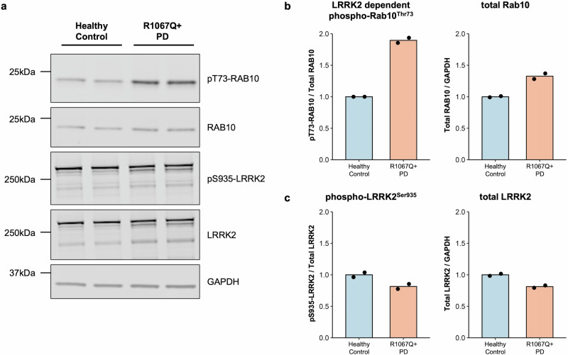
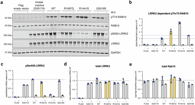
LRRK2-related Parkinson's disease (LRRK2-PD) is the most frequent form of monogenic PD worldwide, with important therapeutic opportunities, exemplified by the advancement in LRRK2 kinase inhibition studies/trials. However, many LRRK2 variants, especially those found in underrepresented populations, remain classified as variants of uncertain significance (VUS). Leveraging on Malaysian, Singaporean, and mainland Chinese PD datasets (n = 4901), we describe 12 Chinese-ancestry patients harboring the LRRK2 p.Arg1067Gln variant, more than doubling the number of previously reported cases (total n = 23, 87% East Asian, mean age of onset: 53.9 years). We determine that this variant is enriched in East Asian PD patients compared to population controls (OR = 8.0, 95% CI: 3.0-20.9), and provide supportive data for its co-segregation with PD, albeit with incomplete penetrance. Utilizing established experimental workflows, this variant showed increased LRRK2 kinase activity, by ~2-fold compared to wildtype and higher than the p.Gly2019Ser variant. Taken together, p.Arg1067Gln should be reclassified from a VUS to pathogenic for causing LRRK2-PD.


Conflict of interest statement

Competing interests: The authors declare no competing interests.

Figures

Fig. 1
Fig. 1. Family pedigrees of the three probands from mainland China with a positive family history of Parkinson’s disease.
There is partial evidence for co-segregation (with the p.Arg1067Gln variant detected in five individuals affected with PD), however with seemingly incomplete penetrance (the youngest sibling in Family 2 [II:5] being clinically unaffected when assessed at age 63 years). The age of onset is written below each affected individual, where available.
Fig. 2
Fig. 2. LRRK2 kinase hyperactivity in vivo due to the presence of the LRRK2 p.Arg1067Gln (p.R1067Q) variant.
Monocyte lysates were analyzed by quantitative immunoblotting (a). Quantified immunoblotting data are presented as ratios of phospho-Rab10Thr73/total Rab10 and total Rab10/GAPDH (b), and phospho-LRRK2Ser935/total LRRK2 and total LRRK2/GAPDH (c), normalized to the average values obtained from the healthy control. The experiments were performed in duplicates, with each data point representing a technical replicate. LRRK2-dependent Rab10 phosphorylation (phospho-Rab10Thr73) as a readout for LRRK2 kinase activity was increased in the monocytes derived from the patient carrying the p.Arg1067Gln variant compared to the control.
Fig. 3
Fig. 3. LRRK2 kinase activity of the LRRK2 p.Arg1067Gln variant compared to p.Gly2019Ser and p.Arg1441Gly in a cellular overexpression system.
In vitro characterization of the LRRK2 p.Arg1067Gln variant in comparison with the common LRRK2 p.Gly2019Ser and p.Arg1441Gly variants in an established HEK293 overexpression system, followed by LI-COR Odyssey immunoblotting and quantification of LRRK2 kinase activity relative to LRRK2 wildtype (wt) (a). Three independent biological replicate transfection experiments were performed including one where cells were treated with and without the specific LRRK2 kinase inhibitor MLi-2 (200 nM for 1.5 h). Each (-) lane represents a biological replicate. LRRK2-dependent phosphorylation of endogenous Rab10 at threonine 73 (pRab10Thr73) was used as a readout for LRRK2 kinase activity, and the LRRK2-specific small molecule inhibitor MLi-2 to demonstrate LRRK2 kinase dependency of pRab10Thr73 as before. LRRK2 kinase hyperactivation was defined as pRab10Thr73 elevation of 1.5-fold compared to LRRK2 wt as before (blue dotted line) (b). Each datapoint represents a biological replicate experiment. p.Arg1067Gln showed LRRK2 activation of 2.4-fold, p.Gly2019Ser of 1.5-fold, and p.Arg1441Gly of 3.4-fold compared to LRRK2 wildtype (b). Expression levels of biomarker phosphorylation of LRRK2 Ser935 (c) and overexpressed LRRK2 (d) as well as endogenous levels of Rab10 (e).
Fig. 4
Fig. 4. Structural insights into predicted impact of the LRRK2 p.Arg1067Gln variant.
Overview of the inactive LRRK2 monomer (a) with detailed view of the Arg1067Gln residue in the inactive LRRK2 monomer (PDB: 8FO2) (b), LRRK2 bound to Type II inhibitor that stabilizes LRRK2 kinase domain in the inactive conformation (PDB: 8U8A) (c), and LRRK2 bound to Type I inhibitor that stabilizes LRRK2 kinase domain in the active conformation (PDB: 8TZH) (d). The interactions that Arg1067 makes with kinase domain residues Phe1883 (electrostatic backbone and potentially Pi-stacking) and Leu1884 (electrostatic backbone) in the inactive conformation are highlighted (b, c). The interaction that Arg1067 makes with kinase domain residues Glu1882 (electrostatic) in the active conformation is also illustrated (d).

Similar articles

Cited by

  • The Global Landscape of Genetic Variation in Parkinson's disease: Multi-Ancestry Insights into Established Disease Genes and their Translational Relevance.
    Lange LM, Fang ZH, Makarious MB, Kuznetsov N, Brolin KA, Ballard S, Bardien S, Doquenia ML, Heutink P, Houlden H, Iwaki H, Jasaityte S, Jones L, Junker J, Kaiyrzhanov R, Koretsky MJ, Kumar KR; Latin American Research Consortium on the Genetics of Parkinson's Disease (LARGE-PD); Leonard HL, Levine KS, Lim SY, Mencacci NE, Mohamed WMY, Nalls MA, Noyce AJ, Ojha R, Okubadejo NU, Rehman SU, Screven L, Shashkin C, Sopromadze S, Stafford EJ, Tan AH, Tan M, Tavadyan Z, Trinh J, Tserensodnom B, Valente EM, Vitale D, Zharkinbekova N, Lohmann K, Bandres-Ciga S, Blauwendraat C, Singleton A, Morris HR, Klein C; Global Parkinson’s Genetics Program (GP2). Lange LM, et al. medRxiv [Preprint]. 2025 Jul 11:2025.07.08.25330815. doi: 10.1101/2025.07.08.25330815. medRxiv. 2025. PMID: 40672482 Free PMC article. Preprint.

References

    1. Blauwendraat, C., Nalls, M. A. & Singleton, A. B. The genetic architecture of Parkinson’s disease. Lancet Neurol19, 170–178, 10.1016/s1474-4422(19)30287-x (2020). - PMC - PubMed
    1. Lim, S. Y. & Klein, C. Parkinson’s disease is predominantly a genetic disease. J. Parkinsons Dis. 10.3233/jpd-230376 (2024). - PMC - PubMed
    1. Lim, S. Y. et al. Uncovering the genetic basis of Parkinson’s disease globally: from discoveries to the clinic. Lancet Neurol. 10.1016/s1474-4422(24)00378-8 (2024). - PubMed
    1. Mata, I. et al. LRRK2: Genetic mechanisms vs genetic subtypes. Handb. Clin. Neurol.193, 133–154, 10.1016/b978-0-323-85555-6.00018-7 (2023). - PubMed
    1. Alessi, D. R. & Sammler, E. LRRK2 kinase in Parkinson’s disease. Science360, 36–37, 10.1126/science.aar5683 (2018). - PubMed

LinkOut - more resources