Review
. 2025 Sep;50(9):3970-3983.
doi: 10.1007/s00261-025-04846-7. Epub 2025 Feb 23.

A pictorial essay on cross-sectional imaging findings of pathologies in the second (D2) segment of the duodenum in adults

Affiliations
Review

A pictorial essay on cross-sectional imaging findings of pathologies in the second (D2) segment of the duodenum in adults

Isil Basara Akin et al. Abdom Radiol (NY). 2025 Sep.

Abstract

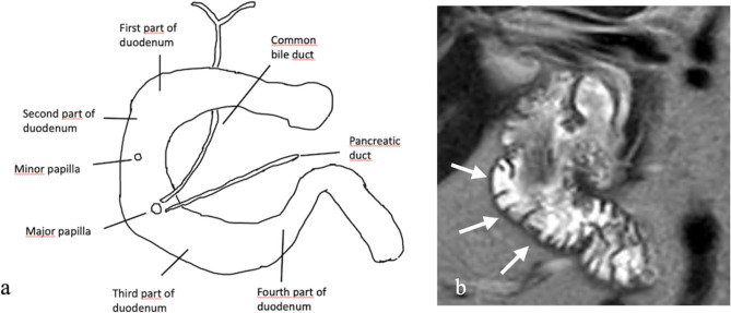
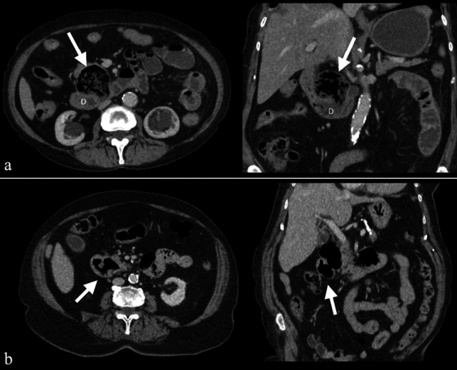
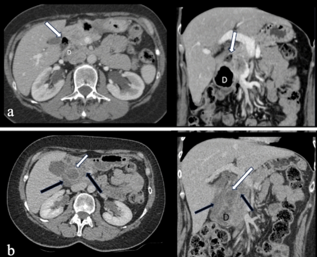
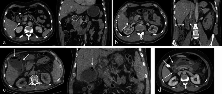
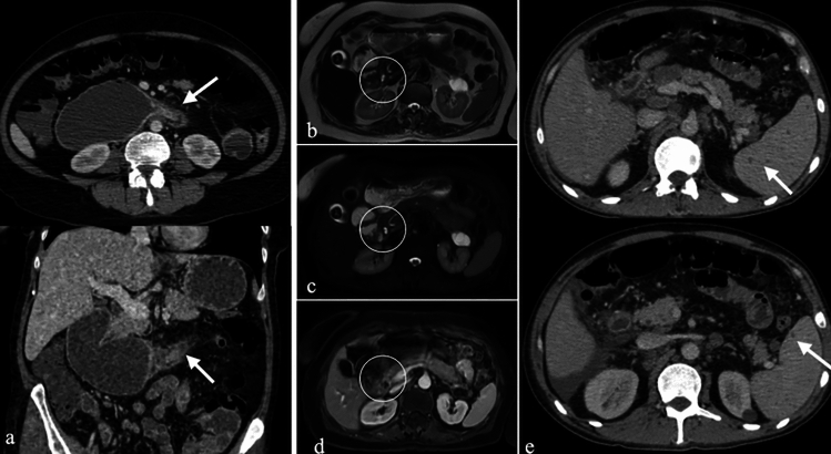
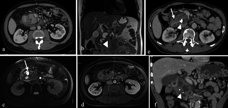
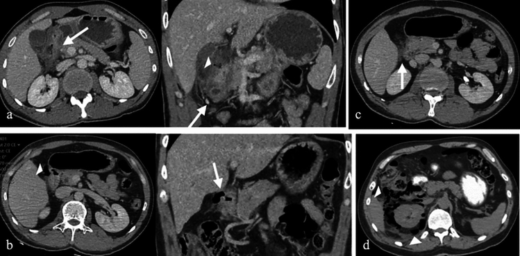
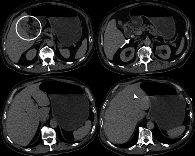
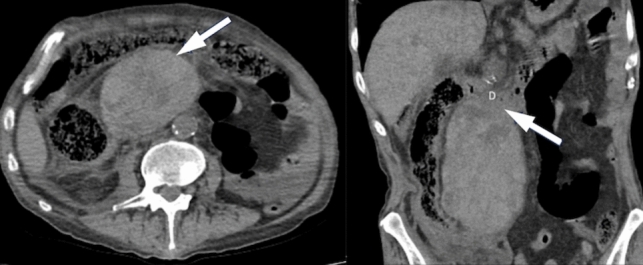
The duodenum, the initial segment of the small intestine, is divided into four parts: the superior (D1), descending (second) (D2), horizontal (D3), and ascending (D4) segments. Despite its short length, the descending part (D2 segment) holds clinical significance due to its anatomical proximity to structures such as the gallbladder, right kidney, colon, and pancreas. This anatomical localization and contiguity give rise to various pathologies, including congenital, inflammatory, infectious, neoplastic, vascular, and traumatic conditions. Cross-sectional imaging modalities play a pivotal role in evaluating pathologies of the second (D2) segment of the duodenum. This article aims to provide a comprehensive overview of these pathologies and delineate their imaging characteristics.

Keywords: Computed tomography; Duodenum; Magnetic resonance imaging; Neoplastic pathologies; Non-neoplastic pathologies.


Conflict of interest statement

Declarations. Conflict of interest: The authors declare no competing interests.

Figures

Fig. 1
Fig. 1
a. Anatomical illustration of the duodenum. b. Coronal T2-weighted MRI image demonstrating the second (D2) segment of the duodenum (arrows)
Fig. 2
Fig. 2
a. Axial and b. Coronal CT images with positive oral and intravenous (IV) contrast administration, c–g MRI images of a 25-year-old male patient with a duodenal duplication cyst, including axial T1-weighted (c), axial T2-weighted (d), coronal T2-weighted (e), coronal post-contrast fat-saturated T1-weighted (f), and MRCP (g) sequences. The cystic lesion exhibits no contrast enhancement. Multiple millimetric hypointense signals, consistent with millimetric stones, are observed on MRI. MRCP reveals no connection with the pancreaticobiliary system (arrows).
Fig. 3
Fig. 3
Axial and coronal negative oral and IV contrast-enhanced CT images of a 57-year-old male patient. A nodular lesion with density and enhancement characteristics similar to pancreatic parenchyma is observed, consistent with ectopic pancreatic tissue (arrows)
Fig. 4
Fig. 4
IV contrast-enhanced a. Coronal and b. Axial CT images. c. Coronal T2-weighted MRI image of a 58-year-old male patient with chronic liver disease. The images demonstrate annular pancreatic tissue encircling the second (D2) segment of the duodenum (arrows), consistent with annular pancreas
Fig. 5
Fig. 5
Axial and coronal IV contrast-enhanced CT images from two different patients (47-year-old male and 53-year-old female) illustrating duodenal diverticula in the second (D2) segment. The diverticulum in the first patient contains intestinal contents (a), while in the second patient, it contains only air (b) (arrows)
Fig. 6
Fig. 6
Axial and coronal IV contrast-enhanced CT images of a 61-year-old female patient. a. CT images obtained five years prior show a duodenal diverticulum (white arrows). b. Recent CT images reveal significant duodenal wall thickening, hyperenhancement, periduodenal heterogeneity, and fluid collections (black arrows), findings consistent with duodenal diverticulitis
Fig. 7
Fig. 7
a. Axial and coronal IV contrast-enhanced CT images of a 43-year-old male patient with peptic ulcer disease, showing discontinuity in the duodenal wall, wall thickening, enhancement, and paraduodenal heterogeneity. b. CT images of a 38-year-old female patient displaying duodenal wall thickening and enhancement, consistent with non-specific duodenitis. c. CT images of a 69-year-old female patient with emphysematous cholecystitis-induced duodenitis, demonstrating air densities within the gallbladder wall. d. CT images of a patient with duodenitis secondary to pancreatitis, revealing non-specific duodenal wall thickening
Fig. 8
Fig. 8
a. Axial and coronal IV contrast-enhanced CT images of a 42-year-old female patient with Crohn’s disease, showing short-segment stenosis, wall thickening, and submucosal fat deposition at the third (D3) segment of the duodenum (arrows), as well as aneurysmal dilatation of the second (D2) segment. MRI images b. T2-weighted, c. fat-saturated T2-weighted, and d. post-contrast fat-saturated T1-weighted) of a 51-year-old female patient with sarcoidosis, showing mucosal thickening and luminal narrowing (circles). e. IV contrast-enhanced CT images of a 68-year-old male patient with duodenal tuberculosis, revealing wall thickening, enhancement, and paraduodenal heterogeneity (arrows), along with intra-abdominal free fluid, splenomegaly, and multiple splenic tuberculous lesions
Fig. 9
Fig. 9
a. Axial IV contrast-enhanced CT, b. Coronal T2-weighted, c. Axial fat-saturated T2-weighted, d. Post-contrast fat-saturated T1-weighted MRI images of a 71-year-old male patient with paraduodenal pancreatitis. Fat stranding and focal fluid collections in the pancreaticoduodenal groove, as well as cystic changes in the duodenal wall, are visible (arrows). Additional findings include dilatation of the common bile duct, choledochus, and pancreatic duct (arrowhead). e. Axial and f. Coronal IV contrast-enhanced CT images of another patient (67-year-old female) with similar findings, along with calcifications within the cystic changes (arrowhead)
Fig. 10
Fig. 10
CT images from four different patients with duodenal perforation: a. Axial and b. Coronal IV contrast-enhanced CT images of a 34-year-old female and a 44-year-old male patient with duodenal ulcer perforations, showing diffuse duodenal wall thickening, periduodenal fat stranding, and free intra-abdominal air (arrows and arrowheads). c. Axial IV contrast-enhanced CT image of a 23-year-old female patient with post-traumatic duodenal perforation. d. Axial IV contrast-enhanced CT image of a 37-year-old male patient with duodenal perforation due to ERCP complications, demonstrating extensive retroperitoneal and periduodenal free air densities (arrowheads)
Fig. 11
Fig. 11
IV contrast-enhanced axial CT images of a 63-year-old female patient with Bouveret’s syndrome. A bilio-duodenal fistula with gallbladder wall thickening and air densities is noted (circle). A large gallstone is impacted in the duodenal bulb (arrow). Gastric dilatation and pneumobilia are evident, consistent with Rigler’s triad
Fig. 12
Fig. 12
Non-enhanced CT images of an 87-year-old female patient, demonstrating an intramural duodenal hematoma affecting the second (D2) and third (D3) segments of the duodenum (arrows)
Fig. 13
Fig. 13
a. Axial and b. Coronal non-enhanced CT images of a 37-year-old male patient with a duodenal lipoma, exhibiting fat attenuation. MRI images c. T1-weighted, d. T2-weighted, and e. fat-saturated T2-weighted of another patient (58-year-old female) showing a lesion with high signal intensity on T1- and T2-weighted images, with signal suppression on fat-saturated sequences (arrows)
Fig. 14
Fig. 14
Axial IV contrast-enhanced CT image of a 39-year-old female patient showing a well-defined, isodense filling defect (arrow), diagnosed as an epithelial polyp
Fig. 15
Fig. 15
a. Non-enhanced axial CT image, b. Axial T2-weighted MRI image of a 62-year-old male patient, showing a round, well-defined, homogeneous lesion diagnosed as leiomyoma
Fig. 16
Fig. 16
Axial IV contrast-enhanced CT images of two patients with duodenal GISTs: a, b A 50-year-old male patient with a well-demarcated hypervascular lesion containing focal calcifications (thick arrow). IV contrast-enhanced c. Axial, d. Coronal CT images of a 67-year-old male patient with a larger lesion containing air densities, indicative of necrosis (thin arrow). e. Axial T1-weighted, b. Axial T2-weighted, c. Axial fat saturated T2-weighted, d. Diffusion weighted, e. ADC maps and f. Contrast enhanced fat saturated T1-weighted MRI images of a 58-year-old female patient diagnosed with GIST, demonstrating hypervascularity, diffusion restriction, and heterogeneous enhancement
Fig. 17
Fig. 17
a. Axial, b. Coronal and c. Sagittal IV contrast-enhanced arterial-phase CT images of a 63-year-old male patient, showing a hypervascular nodular lesion in the second (D2) segment near the major papilla. d. FDG-PET/CT fusion image showing high FDG uptake. e. Coronal T2-weighted MRI image demonstrating hypointensity of the lesion. Diagnosis: neuroendocrine tumor
Fig. 18
Fig. 18
IV contrast enhanced a. Axial, b. Coronal CT, c. FDG-PET CT fusion images and d. Axial T2-weighted e. Coronal T2-weighted f. Diffusion weighted, g. ADC map and h. IV contrast enhanced fat saturated T1-weighted MR images of a 53-year-old female patient with duodenal mass diagnosed as adenocarcinoma. There is concentric wall thickening with heterogeneous enhancement. There is prominent FDG uptake. Lesion has low signal in T2-weighted imaging and shows diffusion restriction. As a result of choledochal involvement, there is dilatation in intrahepatic biller system
Fig. 19
Fig. 19
Non-enhanced CT a. Axial, b. Coronal, c. FDG-PET CT fusion images of a 22-year-old male patient diagnosed as T-cell lymphoma. There is a diffuse segmental wall thickening and prominent FDG uptake compatible with lymphoma involvement of the second—(D2) segment of duodenum. Additionally, there are spheric mesenteric lymph nodes (Arrow)
Fig. 20
Fig. 20
IV contrast enhanced a. Axial and b. Coronal CT images of a 48-year-old male patient with duodenal metastasis of gastric tumor located at cardia. There is an irregular solid mass with necrotic changes and heterogenous enhancement. c. IV contrast enhanced axial CT image of a 52-year-old female patient with colon carcinoma and peritoneal metastasis. In the second—(D2) segment of duodenum there is irregular wall thickening, wall enhancement and paraduodenal fluid collection. Additionally, there are peritoneal nodular lesions, mental heterogeneity and metastatic mass at the fifth segment of liver

Similar articles

References

    1. Reghunath A, Kabilan K, Mittal MK (2020) Exploring the neglected segment of the intestine: the duodenum and its pathologies. Pol J Radiol 85:230-244. 10.5114/pjr.2020.95477 - PMC - PubMed
    1. Ryan S, McNicholas M, Eustace SJ (2024) Anatomy for Diagnostic Imaging. Saunders, Elsevier.
    1. Butler P, Mitchell A, Healy JC et al. (2012) Applied Radiological Anatomy Cambridge University Press.
    1. Gosangi B, Rocha TC, Duran-Mendicuti A (2020) Imaging Spectrum of Duodenal Emergencies. Radiographics 40:1441–1457. 10.1148/rg.2020200045 - PubMed
    1. Fukuhara S, Fukuda S, Sawada H, et al. (2019) A case of duodenal duplication cyst mimicking a pancreatic pseudocyst with intracystic hemorrhage. Surg Case Rep 5:86. 10.1186/s40792-019-0644-3 - PMC - PubMed