

A pictorial essay on cross-sectional imaging findings of pathologies in the second (D2) segment of the duodenum in adults
- PMID: 39988596
- PMCID: PMC12331787
- DOI: 10.1007/s00261-025-04846-7
A pictorial essay on cross-sectional imaging findings of pathologies in the second (D2) segment of the duodenum in adults
Abstract
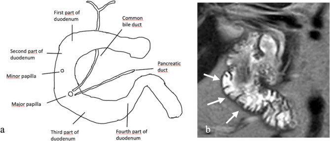
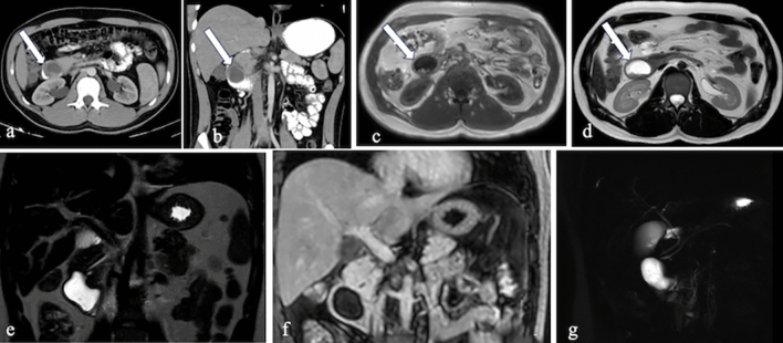
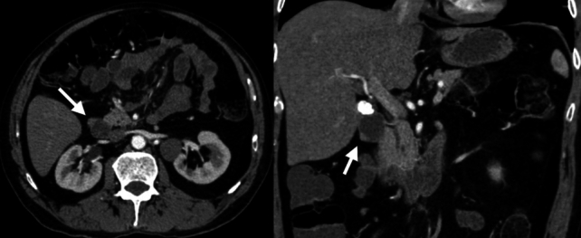
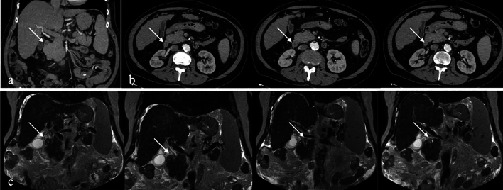
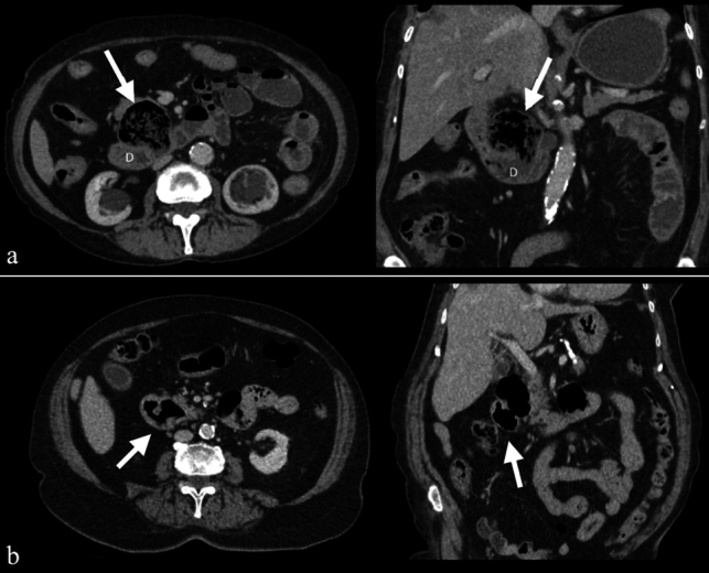
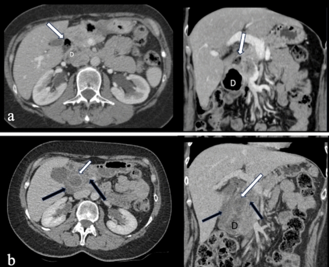
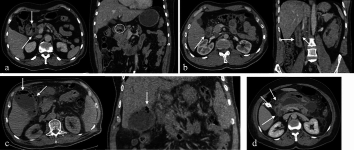
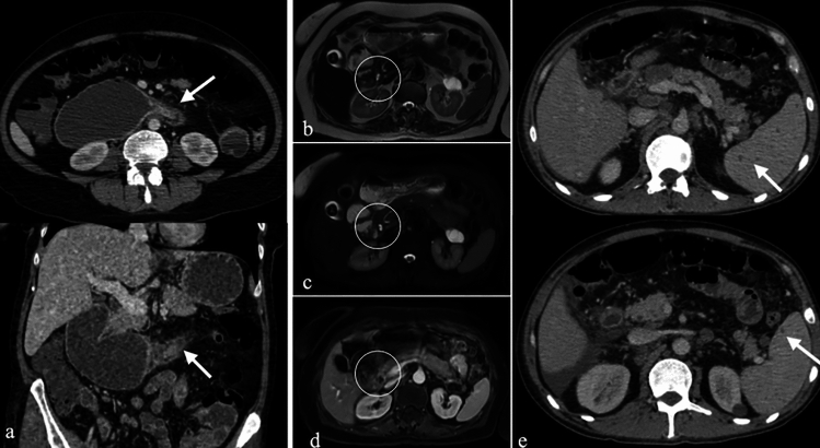
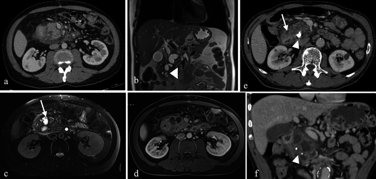
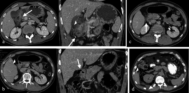
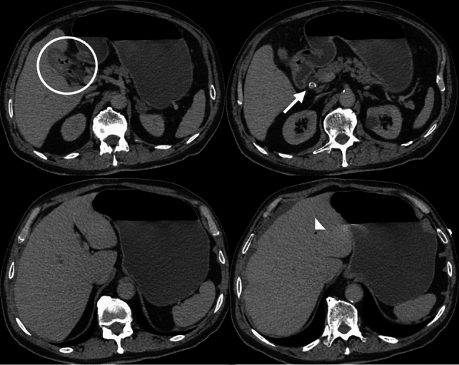
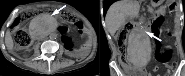
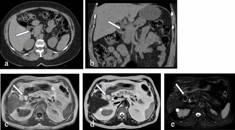
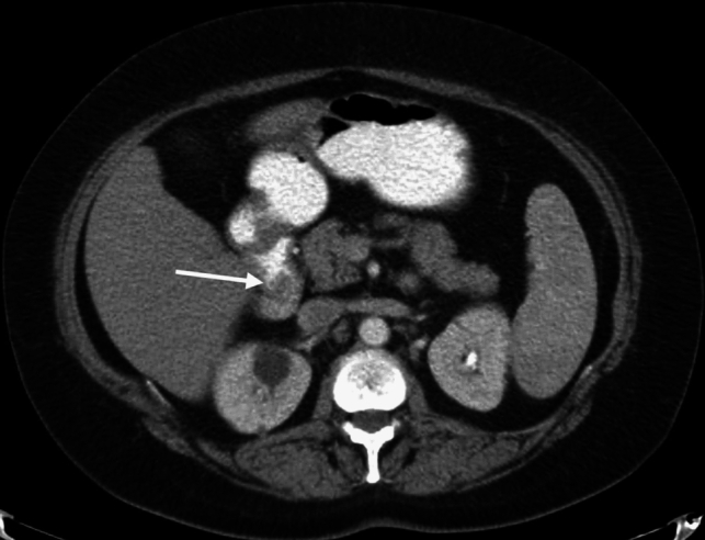
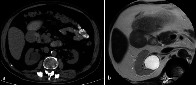
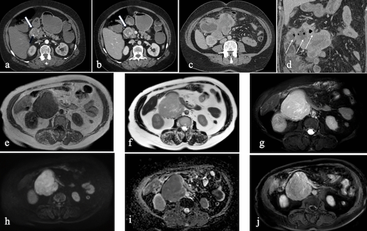
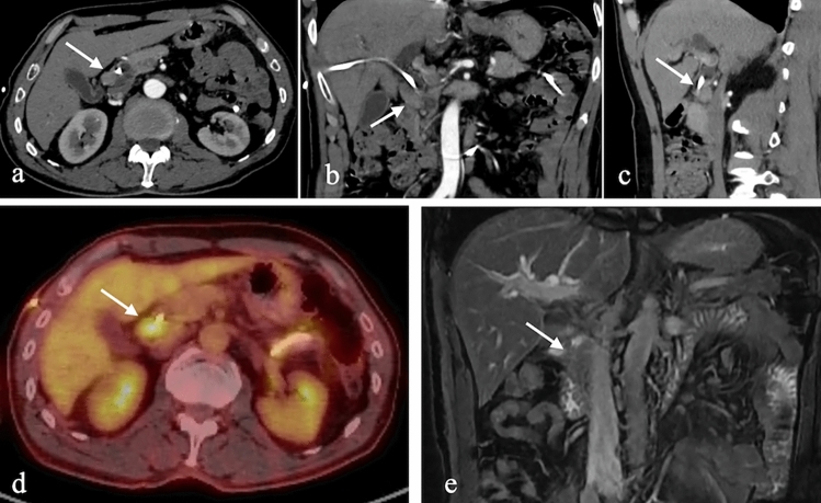
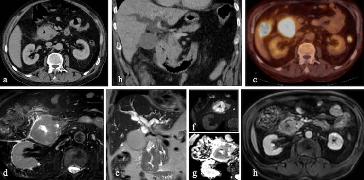
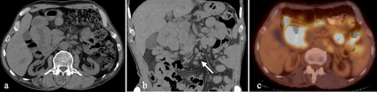
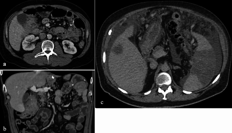
The duodenum, the initial segment of the small intestine, is divided into four parts: the superior (D1), descending (second) (D2), horizontal (D3), and ascending (D4) segments. Despite its short length, the descending part (D2 segment) holds clinical significance due to its anatomical proximity to structures such as the gallbladder, right kidney, colon, and pancreas. This anatomical localization and contiguity give rise to various pathologies, including congenital, inflammatory, infectious, neoplastic, vascular, and traumatic conditions. Cross-sectional imaging modalities play a pivotal role in evaluating pathologies of the second (D2) segment of the duodenum. This article aims to provide a comprehensive overview of these pathologies and delineate their imaging characteristics.
Keywords: Computed tomography; Duodenum; Magnetic resonance imaging; Neoplastic pathologies; Non-neoplastic pathologies.
© 2025. The Author(s).
Conflict of interest statement
Declarations. Conflict of interest: The authors declare no competing interests.
Figures
References
-
- Ryan S, McNicholas M, Eustace SJ (2024) Anatomy for Diagnostic Imaging. Saunders, Elsevier.
-
- Butler P, Mitchell A, Healy JC et al. (2012) Applied Radiological Anatomy Cambridge University Press.
-
- Gosangi B, Rocha TC, Duran-Mendicuti A (2020) Imaging Spectrum of Duodenal Emergencies. Radiographics 40:1441–1457. 10.1148/rg.2020200045 - PubMed
Publication types
MeSH terms
LinkOut - more resources
Full Text Sources
Medical
