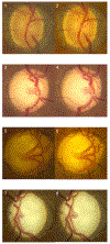
Optic cup morphology associated with glaucomatous damage: Findings from the Primary Open-Angle African American Glaucoma Genetics (POAAGG) Study
- PMID: 39989596
- PMCID: PMC11845224
- DOI: 10.1016/j.ajoint.2024.100053
Optic cup morphology associated with glaucomatous damage: Findings from the Primary Open-Angle African American Glaucoma Genetics (POAAGG) Study
Abstract
Purpose: While it has been well established that advanced glaucoma is associated with a large cup-to-disc ratio (CDR), it is not known if the shape of the cup, particularly excavated cups with expanded lower portions (bean-pot cups), play any additional role in glaucomatous damage. We investigated this among individuals of African ancestry, a population that is more vulnerable to glaucoma than any other ethnic group.
Design: Case-control study.
Methods: Setting:: Institutional (University of Pennsylvania)Subjects:: 3,255 eyes from 1,734 glaucoma cases from the Primary Open-Angle African American Glaucoma Genetics (POAAGG) study.Procedure:: Two graders independently assessed quantitative and qualitative aspects of the optic cup, with any discrepancies adjudicated by an ophthalmologist. The predominant cup shape (>50%) was chosen in cases in which features of two or more cup shapes were present in the same eye. Comparisons of demographic and ocular characteristics among three cup shape groups (conical, cylindrical, and bean-pot) performed using generalized linear models, and generalized estimated equations applied to account for inter-eye correlation.Main Outcome Measures:: Qualitative features of cup shape and phenotypic traits, in conjunction with demographic and genetic information.
Results: Of 3,255 eyes, a total of 1,339 (41.1%) exhibit a conical cup shape, 1,470 (45.2%) have a cylindrical cup shape, and 446 (13.7%) display a bean-pot cup shape. Compared to other cup morphology, bean-pot cups are significantly associated with lower MD, larger CDR, higher IOP, thinner RNFL, and worse VA in logMAR (all p<0.001). Genetic analysis does not show any association between various genetic variants and cup shape. Factors independently predictive of bean-pot cupping include younger age at diagnosis (aOR 0.96 per 1 year increase in age of enrollment, p<0.0001), CDR (adjusted odds ratio (aOR) 1.87, p<0.0001), and the presence of certain optic disc features, including visible pores in the LC (aOR 2.76, p<0.0001), nasalization of vessels (aOR 2.64, p<0.0001), and vessel bayonetting (aOR 2.94, p<0.0001).
Conclusion: This study shows the clinical significance of different cup shapes in glaucoma in an African ancestry population and suggests that bean-pot cups are associated with the most severe glaucomatous damage, independent of cup-disc ratio. This association should be taken into account while determining prognosis following glaucoma interventions.
Figures


References
Grants and funding
LinkOut - more resources
Full Text Sources
