. 2024 Aug 10;14(1):103180.
doi: 10.1016/j.eats.2024.103180. eCollection 2025 Jan.

Achilles Rupture Repair: Modified Gift-Box With a Proximal Myotendinous Backup Fixation Technique

Affiliations

Achilles Rupture Repair: Modified Gift-Box With a Proximal Myotendinous Backup Fixation Technique

Nicholas D Cominos et al. Arthrosc Tech. .

Abstract

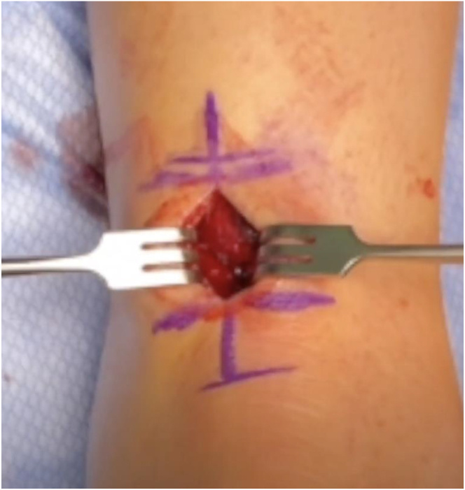
Achilles tendon tears are not an uncommon injury, with a predominance in explosive athletes and weekend warriors in the third to fifth decade of life. Consideration of operative intervention is commonplace in young athletes, whereas less active and older individuals may opt for nonsurgical treatment. Surgical treatment is reported to improve functional outcomes in high-demand individuals and demonstrates increased plantarflexion power, better return to sports rates, and a reduced rerupture rate. Traditionally, a primary end-to-end repair of the Achilles tendon was the surgical treatment of choice. More recently, alternative advanced techniques and minimally invasive constructs have been proposed in an effort to improve repair construct while reducing soft tissue complications. This technical note describes a technique that combines a modified gift-box primary repair with backup fixation using calcaneal anchors. This technique is performed with a small, medially based incision that reduces wound and nerve complications, while promoting end-to-end tendon healing by reducing tension across the repair site through the calcaneal backup fixation.


Conflict of interest statement

The authors (N.D.C., J.S.T., A.J.P., M.A.Z., E.B.E, L.S.K.) declare that they have no known competing financial interests or personal relationships that could have appeared to influence the work reported in this paper.

Figures

Fig 1
Fig 1
The patient is positioned prone, with the left ankle joint at the end of the bed to allow the limb to lay flat. The palpable gap localizing the Achilles tendon rupture site is identified (arrow) as the distal extent of the planned surgical incision.
Fig 2
Fig 2
The patient is prone and the posterior aspect of the ankle is shown, centered over the achilles tendon. A small 2- to 4-cm open incision is used to minimize insult to soft tissues but also to allow proper visualization for repair and avoidance of neurovascular structures. This incision is cheated medially to reduce risk to the sural nerve. The distal extent of the incision is planned at the previously identified palpable gap of the rupture site.
Fig 3
Fig 3
The patient is prone and the medial ankle is visualized. The proximal tendon stump is delivered into the wound, and a tongue depressor can be used as a base to allow for safe, sharp dissection of devitalized tissue (arrow).
Fig 4
Fig 4
The patient is prone and the achilles tear is exposed. The proximal end of the tendon is secured with Krackow locking repair sutures exiting the face of the tear (arrow). Additionally, a locking suture tape is separately passed proximal to the repair suture, exiting deep to the tendon, which will be used for backup fixation into the calcaneus (arrowhead).
Fig 5
Fig 5
The patient is prone and the ankle is resting on a bump to aid in positioning of the distal achilles stump. A straight suture lasso is passed into the calcaneal stab incision (arrow) and through the distal Achilles tendon, exiting at the face of the torn tendon. Palpation of the end of the torn tendon can help localize for accurate lasso passage. This will be used to pass the deep suture tapes through the distal tendon for eventual backup fixation into the calcaneus.
Fig 6
Fig 6
The patient is prone and the medial ankle is visualized. The modified gift-box repair is started by passing the micro lasso through the medial side of the distal tendon stump (arrow), exiting at the face of the tear and aiming laterally. The suture securing the lateral aspect of the proximal tendon stump (arrowhead) will be loaded into the passing wire and pass through the distal Achilles tendon. This will be repeated laterally on the distal stump, as well as on both sides of the proximal aspect of the tear.
Fig 7
Fig 7
The patient is prone with the medial aspect of the ankle visualized. Final Achilles tendon repair construct. The modified gift box serves to secure the tendon ends to one another, without tying knots directly at the repair site (arrow). Additionally suture anchors into the calcaneus provide strong backup fixation and take tension of the repair site.

References

    1. Forlenza E.M., Lavoie-Gagne O.Z., Lu Y., Diaz C.C., Chahla J., Forsythe B. Return to play and player performance after Achilles tendon rupture in UEFA professional soccer players: A matched-cohort analysis of players from 1999 to 2018. Orthop J Sports Med. 2021;9 - PMC - PubMed
    1. Deng S., Sun Z., Zhang C., Chen G., Li J. Surgical Treatment Versus Conservative Management for Acute Achilles Tendon Rupture: A systematic review and meta-analysis of randomized controlled trials. J Foot Ankle Surg. 2017;56:1236–1243. - PubMed
    1. Cretnik A., Kosir R. Incidence of Achilles tendon rupture: 25-year regional analysis with a focus on bilateral ruptures. J Int Med Res. 2023;51 - PMC - PubMed
    1. Ho G., Tantigate D., Kirschenbaum J., Greisberg J.K., Vosseller J.T. Increasing age in Achilles rupture patients over time. Injury. 2017;48:1701–1709. - PubMed
    1. Shamrock A.G., Dreyer M.A., Varacallo M. StatPearls; FL: 2024. Achilles tendon rupture. Treasure Island. - PubMed

LinkOut - more resources