Review
. 2025 Feb 4;10(6):5214-5250.
doi: 10.1021/acsomega.4c09806. eCollection 2025 Feb 18.

Advancements in Micro/Nanorobots in Medicine: Design, Actuation, and Transformative Application

Affiliations
Review

Advancements in Micro/Nanorobots in Medicine: Design, Actuation, and Transformative Application

Induni Nayodhara Weerarathna et al. ACS Omega. .

Abstract

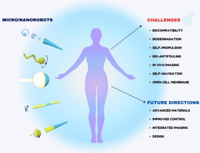
In light of the ongoing technological transformation, embracing advancements that foster shared benefits is essential. Nanorobots, a breakthrough within nanotechnology, have demonstrated significant potential in fields such as medicine, where diagnostic and therapeutic applications are the primary focus areas. This review provides a comprehensive overview of nanotechnology, robots, and their evolving role in medical applications, particularly highlighting the use of nanorobots. Various design strategies and operational principles, including sensors, actuators, and nanocontrollers, are discussed based on prior research. Key nanorobot medical applications include biomedical imaging, biosensing, minimally invasive surgery, and targeted drug delivery, each utilizing advanced actuation technologies to enhance precision. The paper further examines recent progress in micro/nanorobot actuation and addresses important considerations for the future, including biocompatibility, control, navigation, delivery, targeting, safety, and ethical implications. This review offers a holistic perspective on how nanorobots can reshape medical practices, paving the way for precision medicine and improved patient outcomes.


Conflict of interest statement

The authors declare no competing financial interest.

Figures

Figure 1
Figure 1
Schematic diagram of fabrication methods of micro/nanorobots.
Figure 2
Figure 2
A) CoPt nanowires are synthesized using a template-assisted electrodeposition process. B) The magnetization angle of hard-magnetic CoPt nanowires aligns with the short axis (yellow). In contrast, soft-magnetic CoNi nanowires align with the direction of the magnetic field (red). Reproduced with permission from the ref Copyright 2019 American Chemical Society. C) The dumbbell-shaped MagRobot is composed of a nickel nanowire (Ni NW) connected to two polystyrene (PS) microbeads. Reproduced with permission from ref (48). Copyright 2016 Wiley-VCH Verlag GmbH and Co. KGaA, Weinheim. D) A fish-like nano swimmer exhibits traveling-wave motion when subjected to an oscillating magnetic field. Reproduced with permission from ref (49). Copyright 2016 Wiley-VCH Verlag GmbH and Co. KGaA, Weinheim. E) A two-arm nano swimmer can freestyle swimming. Reproduced with permission from ref (50). Copyright 2017 American Chemical Society. F) SEM images show 1-, 2-, and 3-link microswimmers, with the 3-link microswimmer demonstrating traveling-wave propulsion under an oscillating magnetic field. Reproduced with permission from ref (51). Copyright 2015 American Chemical Society. G) PVDF-Ppy-Ni nanoeels exhibit three distinct motion modes, as illustrated in the SEM image provided. Reproduced with permission from ref (52). Copyright 2019 Wiley-VCH Verlag GmbH and Co. KGaA, Weinheim.
Figure 3
Figure 3
(A) Fe-coated camptothecin-loaded magnetic tube for killing HeLa cells. White circles highlight dead cells. (B) Controllable navigation and targeted transport of antibodies inside blood flow by using Janus micropropellers. (C) Sperm-based MagRobots are capable of delivering heparin-loaded liposomes through flowing blood. (D) pDNA transfection by human embryo kidney cells when in targeted contact with helical microrobots loaded with plasmid DNA. (E) Released drugs from hydrogel-based microswimmer for active labeling.
Figure 4
Figure 4
(A) Schematic process of removing cholesterol plaque in the blood artery via the magnetic hyperthermia of nanorobots. (B) Experimental setup of Janus nanorobots for magnetically induced thermophoresis. Thermophoretic force, triggered by the temperature difference, causes the self-propulsion of a Janus particle. (C) Underlying physics of the magnetoelectrically triggered drug (i.e., AZTTP) release process.
Figure 5
Figure 5
(A) The structural similarities and differences between natural and artificial muscles. (Adapted with permission from ref (132). Copyright 2021, Microsystems and Nanoengineering.) (B) Polarized optical microscope of liquid crystal formation observed at 45° angles concerning cross-polarizers (top) and scanning electron microscope image of an electrographite flake (bottom). (C) The shape deformation of artificial muscles during relaxation and contraction. (Adapted with permission from ref (133). Copyright 2022, Springer Nature.)
Figure 6
Figure 6
A) A controlled, needle-like LMGNS that moves when exposed to NIR laser. Reproduced with permission. Copyright 2021, Elsevier. B) B-TiO2/Ag nanorobot fabrication and characterization. Reproduced with permission. Copyright 2022, Wiley-VCH. C) Schematic of The Pt NP-modified polyelectrolyte multilayer manufacturing process of micro engines. Reproduced with permission. Copyright 2014, American Chemical Society. D) Schematic illustration of DOX-functionalized carboxy betaine microrobots for UV-light-triggered drug release and an example for the drug release demonstration. Reproduced with permission. Copyright 2020, Wiley-VCH.
Figure 7
Figure 7
A) Convert porphyrin nanodroplets into PFC microbubbles by phase transition. Used by permission. Copyright Wiley-VCH, 2016. B) A PFC-powered microcannon that fires NBs. Used by permission. Copyright The American Chemical Society. All rights reserved. C) Experimental design of fluorescein isothiocyanate-labeled mesoporous silica nanoparticle delivery via ultrasound-mediated cavitation-enhanced extravasation in the agarose phantom model. Used by permission. Copyright Elsevier 2018.
Figure 8
Figure 8
A). The urease micro- and nanomotors’ AMP-coating technique allows for independent propulsion of the motors, which can target pathogenic infections in vivo and in vitro. Used by permission. The American Chemical Society. Copyright 2022. B) Au–Pt bimetallic nanorod in an aqueous H2O2 solution driven by a self-electrophoresis process. Used by permission. All rights reserved by Wiley-VCH, 2018. C) Diagram showing the catalytic helical carbon motor system’s propulsion. Used by permission. D) Diagrammatic representation of the production processes for reversed Janus motors and the different mobility scenarios. Copyright 2019, Wiley-VCH. Used by permission. The American Chemical Society. All rights reserved. E) SMMC and SMMF optical pictures show how pH variations cause the fins and claws to open and close. Reproduced with permission. Copyright 2021, American Chemical Society.
Figure 9
Figure 9
(a) Schematic illustration of magnetically navigated, ultrasonically propelled RBC micromotors in the whole blood. Reproduced with permission from reference (165). Copyright 2014, American Chemical Society. (b) Sperm flagella-driven microbiorobots. Reproduced with permission from reference (162). Copyright 2013, Wiley-VCH Verlag GmbH & Co. KGaA, Weinheim. (c) Bacteria-driven microswimmers based on PEM-MNP microparticles attached to E. coli MG1655 bacteria. Reproduced with permission from reference (166). Copyright 2017, American Chemical Society. (d) Magnetically guided, bacterially driven RBC microswimmers for active drug delivery. Reproduced with permission from reference (167). Copyright 2018, Science Robotics.
Figure 10
Figure 10
Diagram showing how conventional drugs were supplied without using nanocarriers, causing damage to cells or organs that were considered healthy. By contrast, current techniques use nanorobots to deliver drugs to targeted body areas.
Figure 11
Figure 11
(A) Zinc-based micromotors driven by acid for improved retention in the mouse stomach. (Adapted with permission from ref (172). Copyright 2015, American Chemical Society.) (B) To avoid the acidic stomach environment and instead selectively position and propel spontaneously within the gastrointestinal tract, an enteric micromotor coated with a pH-sensitive polymer barrier (enteric coating) is used. (Adapted with permission from ref (173). Copyright 2016, American Chemical Society.) (C) Regulated swimming of a swarm of microrobotic flagella that resemble bacteria in vivo (Adapted with permission from ref (174). Copyright 2015, John and Wiley.) (D) Drug-containing nanoliposomes are delivered to tumor-hypoxic areas by magneto-aerotactic motor-like bacteria. (Adapted with permission from ref (175). Copyright 2016, Springer Nature.)
Figure 12
Figure 12
(A) Macrophage-magnesium biohybrid micromotor. (Adapted with permission from ref (176). Copyright 2019 John Wiley and Sons.) (B) Bacteriophage virus-like nanoparticles (QβVLPs)-loaded Mg-based micromotors. (Adapted with permission from ref (177). Copyright 2020, John Wiley and Sons.) (C) Mg-Fe3O4-based magneto-fluorescent nanorobot. (Adapted with permission from ref (178). Copyright 2021, Springer Nature.) (D) Poly(aspartic acid)/iron–zinc microrockets. (Adapted with permission from ref (179). Copyright 2019, American Chemical Society.) (E) Calcium carbonate micromotors. (Adapted with permission from ref (180). Copyright 2016, Springer Nature.) (F) NO-driven nanomotors (Adapted with permission from ref (119). Copyright 2019, Springer Nature.)
Figure 13
Figure 13
(A) Schematic and experimental images (inset) depict rolled-up magnetic microdrillers with a sharp end penetrating a pig liver postdrilling motion. (B) Schematic of a driller operating in a 3D vascular network and experimental results showing the driller dislodging a blood clot. (C) The movement of an Au/Ag/Ni surface walker under varying frequencies of a transversal rotating field shows the magnetic navigation of microrobots penetrating and removing a cell fragment. (D) Magnetic manipulation of Si/Ni/Au nanospheres for targeted intracellular transfection. (E) Penetration of Helicobacter pylori, a helical MagRobot, into mucin gels and mucus liquefaction via enzyme-catalyzed reaction. (F) Long-range propulsion of injected slippery MagRobots in the vitreous toward the retina, aided by a magnetic field and standard optical coherence tomography.
Figure 14
Figure 14
(A) MRI of microrobot swarms in rat stomach tissue after magnetic actuation over time. Reproduced with permission from ref (169). Copyright 2017, Science Robotics. (B) The image-guided theranostic platform combining magnetic particle imaging and localized hyperthermia in a U87MG xenograft mouse shows superparamagnetic nano robots in the liver and tumor. Reproduced with permission from ref (212). Copyright 2018 ACS. (C) In vivo fluorescence of spirulina-based MagRobots in mouse intraperitoneal cavity over time. Reproduced with permission from ref (169). Copyright 2017, Science Robotics. (D) Ultrasound tracking of MagRobot swarm generation in a bovine eyeball. Reproduced with permission from ref (213). Copyright 2019 Nature. (E) Real-time tracking of microrobots in phantoms using multispectral optoacoustic tomography actuated by a permanent magnet. Reproduced with permission from ref (214). Copyright 2019 ACS. (F) SPECT images of radiolabeled microrobots in an Eppendorf tube and mice. Reproduced with permission from ref (215). Copyright 2019 Wiley-VCH.
Figure 15
Figure 15
(A) Equipping micro/nano robots with various bioreceptors to enable them to sense target analytes, such as proteins, nucleic acids, and cells. (B) Microrockets functionalized with ssDNA for nucleic acid separation and selective hybridization. (Adapted with permission from ref (246). Copyright 2011 American Chemical Society.) (C) Ultrasound-propelled nanomotors for the specific intracellular detection of miRNA in intact cancer cells. (Adapted with permission from ref (247). Copyright 2015 American Chemical Society.)
Figure 16
Figure 16
Limitations and future directions of micro/nanorobots

Similar articles

Cited by

References

    1. Jain K. Advances in the field of nanooncology. BMC Med. 2010, 8, 83.10.1186/1741-7015-8-83. - DOI - PMC - PubMed
    1. Misra R.; Acharya S.; Sahoo S. K. Cancer nanotechnology: application of nanotechnology in cancer therapy. Drug Discovery Today. 2010, 15, 842–50. 10.1016/j.drudis.2010.08.006. - DOI - PubMed
    1. Rhyne P. W.; Wong O. T.; Zhang Y. J.; Weiner R. S. Electrochemiluminescence in bioanalysis. Bioanalysis. 2009, 1, 919–35. 10.4155/bio.09.80. - DOI - PubMed
    1. Matsue T. Bioimaging with Micro/Nanoelectrode Systems. ANAL SCI. 2013, 29, 171–9. 10.2116/analsci.29.171. - DOI - PubMed
    1. Choi Y.-E.; Kwak J.-W.; Park J. W. Nanotechnology for Early Cancer Detection. Sensors. 2010, 10, 428–55. 10.3390/s100100428. - DOI - PMC - PubMed

LinkOut - more resources