[Preprint]. 2025 Feb 13:rs.3.rs-5876218.
doi: 10.21203/rs.3.rs-5876218/v1.

Crystallographic fragment screening and deep mutational scanning of Zika virus NS2B-NS3 protease enable development of resistance-resilient inhibitors

Affiliations

Crystallographic fragment screening and deep mutational scanning of Zika virus NS2B-NS3 protease enable development of resistance-resilient inhibitors

Frank von Delft et al. Res Sq. .

Abstract

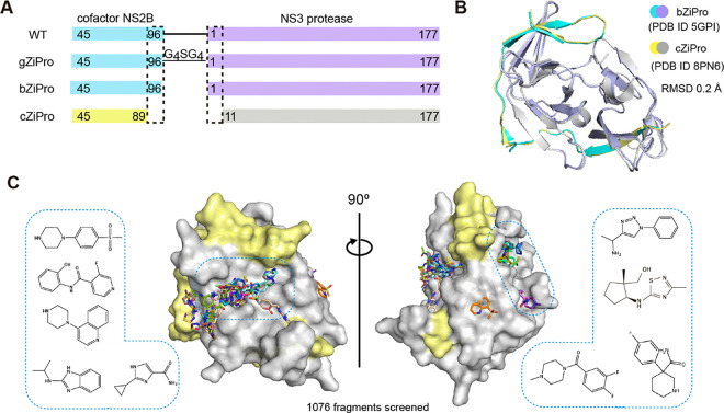
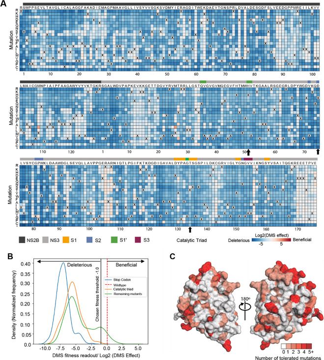
The Zika viral protease NS2B-NS3 is essential for the cleavage of viral polyprotein precursor into individual structural and non-structural (NS) proteins and is therefore an attractive drug target. Generation of a robust crystal system of co-expressed NS2B-NS3 protease has enabled us to perform a crystallographic fragment screening campaign with 1076 fragments. 47 fragments with diverse scaffolds were identified to bind in the active site of the protease, with another 6 fragments observed in a potential allosteric site. To identify binding sites that are intolerant to mutation and thus suppress the outgrowth of viruses resistant to inhibitors developed from bound fragments, we performed deep mutational scanning of NS2B-NS3 protease. Merging fragment hits yields an extensive set of 'mergers', defined as synthetically accessible compounds that recapitulate constellations of observed fragment-protein interactions. In addition, the highly sociable fragment hits enable rapid exploration of chemical space via algorithmic calculation and thus yield diverse possible starting points that maximally explore the binding opportunities to NS2B-NS3 protease, facilitating its resistance-resilient antiviral development.

Keywords: NS2B-NS3 protease; crystallographic fragment screening; deep mutational scanning; sociable fragments.


Conflict of interest statement

Competing Interest Statement A.S.G consults for DNDi and MMV.

Figures

Figure 1:
Figure 1:. Crystallographic fragment screening of ZIKV NS2B-NS3 protease.
(A) Domain boundaries of constructs gZiPro, bZiPro and cZiPro. (B) Structure superimposition of bZiPro structure 5GPI and cZiPro structure 8PN6 with a RMSD of 0.2 Å. (C) Surface view of ZIKV NS2B-NS3 fragment screening output in two orientations. NS3 protein surface is coloured in grey and NS2B is coloured in yellow. Identified fragments are shown as sticks. Examples of observed fragments are listed in dashed box.
Figure 2:
Figure 2:. Interaction motifs for ligand engagement in the active site of ZIKV NS2B-NS3 protease.
(A) Surface view of observed fragments bound in the active site. The active site is divided into subsites S1, S1’, S2 and S3 based on substrate residue binding position. Representative examples of fragments Z336080990(x0089) (B), Z396117078(x1098) (C) and Z425338146(x0404) (D) interacting at the S1, S1-S1’, and S2 site, respectively. The PanDDA event map is shown as a dark grey mesh. Hydrogen bonds are shown as dashed lines. (E)The unique fragment Z1587220559(x0846b) observed in S1’ site. (F) A plot of key residues observed for fragment interaction. Y-axis represents the number of fragments forming interaction with associated residues. (G) Key interacting residues revealed from fragment screening. HBA: hydrogen bond acceptor. HBD: hydrogen bond donor.
Figure 3:
Figure 3:. Deep mutational scanning to measure mutational tolerance of ZIKV NS2B-NS3.
(A) The heatmap of NS2B-NS3 residues indicating the mutational effect of each amino acid substitutions in the ZIKV NS2B-NS3 protease. Blue mutations are deleterious for viral growth in Huh-7.5 cells relative to wild-type, white mutations are neutral, and red mutations increase growth in Huh-7.5 cells. Wildtype amino-acid identities at each site are denoted by an ‘X’. In general, most mutations decrease the fitness of the virus in Huh-7.5 cells. (B) Distribution of fitness estimates for all mutations. The distributions of tness effects of stop-codon mutants, Catalytic triad mutants and the remaining mutants are coloured in blue, orange and green, respectively. The black vertical line indicates the threshold −1.0 (Log2 (DMS effect)) for fitness. (C) Fitness view of NS2B-NS3 protease. Mutational tolerance is mapped onto the structure of ZIKV NS2B-NS3 (PDB ID: 8PN6). Residues marked in white do not tolerate changes to that site, while residues marked in red tolerate a range of changes. The number of changes tolerated is indicated by the number and the colour.
Figure 4:
Figure 4:. Fitness view reveals potential mutational effect on ligand engagement in the active site.
(A) Fitness view of the active site with fragment Z396117078(x1098) presented. Key interacting residues identified from fragment screening are numbered. Mutational intolerant region is coloured in white, and mutational tolerant region is coloured in red. (B) Logo plot of experimentally measured amino acid preferences of the label key interacting residues in figure A. High mutational residues are marked with an asterisk sign. (C) Example of Fragment Z1203191681(x0719) formed a direct H-bond with the hydroxyl group of tyrosine 161. (D) Key interacting residues in the active site. Regions that suggest to form backbone interacting are coloured in blue. Side chain of Y161 is highlighted by colour orange due to its potential mutation to phenylalanine. Opportunity for fragment growth is shown as an arrow.
Figure 5:
Figure 5:. Fragments bound in a non-active site of ZIKV NS2B-NS3 protease.
(A) Overview of fragments observed in the non-active site. (B) Structural details of observed fragment Z57122377(x0130) interacting at AS1’. Residues that participate in fragment interaction are shown as sticks. Representative examples of fragment Z1428159350 (x0806) (C) and Z1272517105 (x0777) (D) bound in the AS2’ site. Hydrogen bonds are shown as dashed lines. The PanDDA event map is shown as a dark grey mesh. (E) Surface view of an allosteric site defined by reported docking and mutational studies. Labelled residues are reported for ligand interaction. Oxygen, nitrogen and carbon atoms are coloured in red, blue and cyan, respectively. (F) Fitness view of the potential allosteric site mapping to Figure E. The logo plot presents the experimentally measured amino acid preferences of the interacting residues in the non-active site. High mutational residues are marked with an asterisk sign.
Figure 6:
Figure 6:. Screened crystallographic fragments tested in CreoptixWAVE binding assay and inhibitory activity assay.
Fragment Z270834034(x0472) (A) and Fragment Z1269184613(x0772) (B) showed close to mM affinity, but such results could not be reproduced. (C) Fragment Z1587220559(x0846) selected as an example of negative result. (D) Peptide-hybrid inhibitor compound 36 was used as the positive control, showing a Kd of 0.89 μM in the GCI assay. Compound 36 was tested at 1 μM, while fragments were tested at 250 μM. 2D structures of compounds are shown on top. (E-H) Fluorescence-dose-response inhibitory activity assay. Fragments Z270834034(x0472) (E), Z1269184613(x0772) (F) and Z1587220559(x0772) (G) showed no inhibitory activity. (H) Positive control compound 36 showing an IC50 of 1.8 μM.
Figure 7:
Figure 7:. Fragment bridging multiple sites providing opportunities for merging and linking.
(A) Fragments selected as parent hits for rapid follow-up compound design. (B) Fragment alignment shows opportunities for merging and linking. (C) Predicted mergers posed in the active site. 2D structure is shown at top with its Enamine compound identity code. RMSD value indicates the conformational variance to its parent fragments. (D) An annotated plot of the chemical diversity of 4,000 filtered catalogue compounds that are close analogues (graph edit distance fewer than 6 edits) of mergers of the active site fragment-hits searched with SmallWorld in Enamine and Mcule catalogues.

References

    1. de Araujo TVB, Ximenes RAA, Miranda-Filho DB, Souza WV, Montarroyos UR, de Melo APL, Valongueiro S, de Albuquerque M, Braga C, Filho SPB et al. (2018) Association between microcephaly, Zika virus infection, and other risk factors in Brazil: final report of a case-control study. Lancet Infect Dis 18(3):328–336. 10.1016/S1473-3099(17)30727-2 - DOI - PMC - PubMed
    1. Krauer F, Riesen M, Reveiz L, Oladapo OT, Martinez-Vega R, Porgo TV, Haefiger A, Broutet NJ, Low N, Group WH (2017) O. Z. C. W. Zika Virus Infection as a Cause of Congenital Brain Abnormalities and Guillain-Barre Syndrome: Systematic Review. PLoS Med 14(1):e1002203. 10.1371/journal.pmed.1002203 - DOI - PMC - PubMed
    1. Lowe R, Barcellos C, Brasil P, Cruz OG, Honorio NA, Kuper H, Carvalho MS (2018) The Zika Virus Epidemic in Brazil: From Discovery to Future Implications. Int J Environ Res Public Health 15(1). 10.3390/ijerph15010096 - DOI - PMC - PubMed
    1. Saiz JC, Therapeutic Advances Against ZIKV (2019) A Quick Response, a Long Way to Go. Pharmaceuticals (Basel) 12 (3). DOI: 10.3390/ph12030127. - DOI - PMC - PubMed
    1. Chappell KJ, Stoermer MJ, Fairlie DP, Young PR (2006) Insights to substrate binding and processing by West Nile Virus NS3 protease through combined modeling, protease mutagenesis, and kinetic studies. J Biol Chem 281(50):38448–38458. 10.1074/jbc.M607641200 - DOI - PubMed

Publication types

LinkOut - more resources