[Preprint]. 2025 Feb 11:rs.3.rs-4138879.
doi: 10.21203/rs.3.rs-4138879/v1.

A clinical drug candidate that triggers non-apoptotic cancer cell death

Affiliations

A clinical drug candidate that triggers non-apoptotic cancer cell death

Scott Dixon et al. Res Sq. .

Update in

  • Tegavivint triggers TECR-dependent nonapoptotic cancer cell death.
    Leak L, Wang Z, Joseph AJ, Johnson B, Chan AA, Decosto CM, Magtanong L, Ko PJ, Lee WC, Ritho J, Manukian S, Millner A, Chitkara S, Salinas JJ, Skouta R, Rees MG, Ronan MM, Roth JA, Myers CL, Moffat J, Boone C, Bensinger SJ, Nathanson DA, Atilla-Gokcumen GE, Moding EJ, Dixon SJ. Leak L, et al. Nat Chem Biol. 2025 Dec;21(12):1873-1884. doi: 10.1038/s41589-025-01913-4. Epub 2025 May 26. Nat Chem Biol. 2025. PMID: 40419770

Abstract

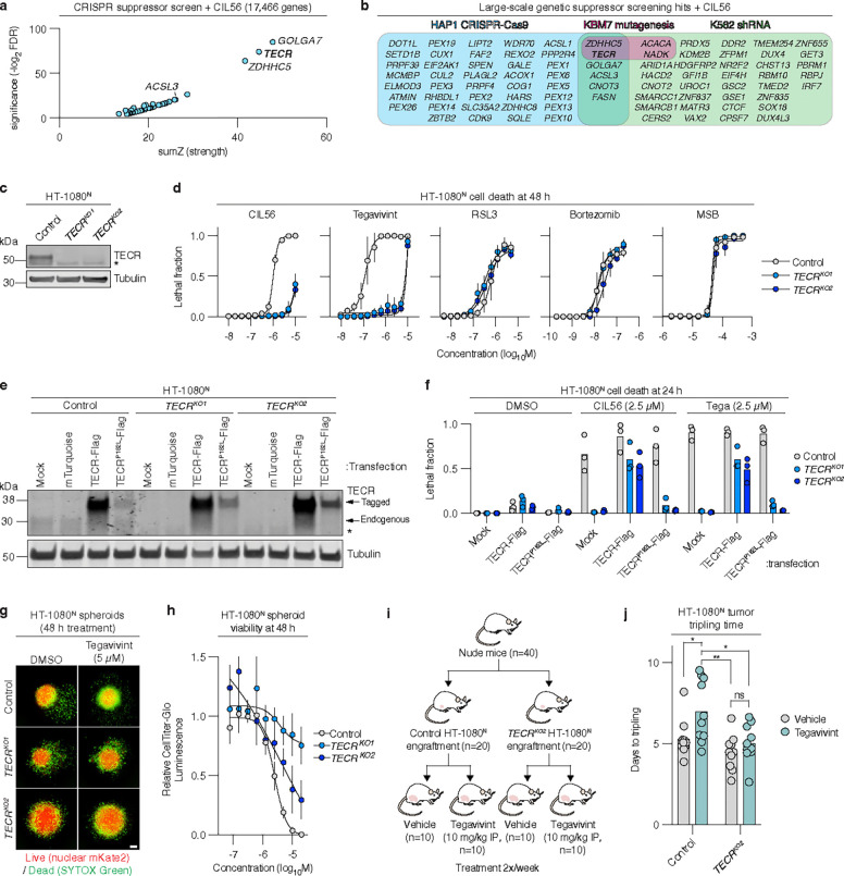
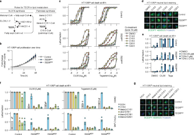
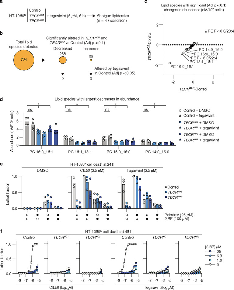
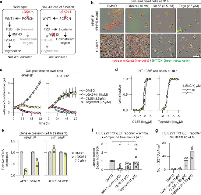
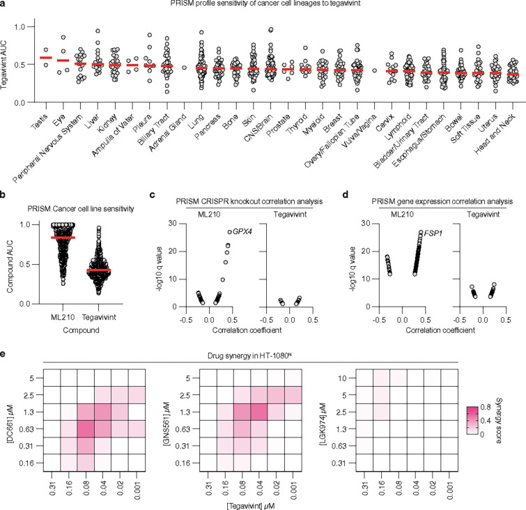
Small molecules that induce non-apoptotic cell death are of fundamental mechanistic interest and may be useful to treat certain cancers. Here, we report that tegavivint, a drug candidate undergoing human clinical trials, can activate a unique mechanism of non-apoptotic cell death in sarcomas and other cancer cells. This lethal mechanism is distinct from ferroptosis, necroptosis and pyroptosis and requires the lipid metabolic enzyme trans-2,3-enoyl-CoA reductase (TECR). TECR is canonically involved in the synthesis of very long chain fatty acids but appears to promote non-apoptotic cell death in response to CIL56 and tegavivint via the synthesis of the saturated long-chain fatty acid palmitate. These findings outline a lipid-dependent non-apoptotic cell death mechanism that can be induced by a drug candidate currently being tested in humans.

Keywords: TECR; cancer; necrosis; palmitate.


Conflict of interest statement

Competing interests statement E.J.M. has served as a paid consultant for Guidepoint and GLG. S.J.D. is an inventor on patents related to ferroptosis. Additional Declarations: Yes there is potential Competing Interest. E.J.M. has served as a paid consultant for Guidepoint and GLG. S.J.D. is an inventor on patents related to ferroptosis.

Figures

Figure 1
Figure 1. Structurally related synthetic oximes can trigger non-apoptotic cell death.
a, Small molecule structures. b, Cell death determined by imaging of live (nuclear mKate2-positive) and dead (SYTOX Green-positive) cells. Live and dead cell counts were integrated into lethal fraction scores, which were then summed across an 11-point 2-fold dose-response (100 μM high concentration). c, Cell death determined by imaging of live and dead cells. A lethal fraction score of 0 = all cells in the population are alive, 1 = all cells in the population are dead. QVD, Q-VD-OPh. d,e, Protein abundance determined by immunoblotting and quantified across three independent experiments. Compound treatments were EC90 values, with samples harvested right before the onset of cell death: CIL56 (1.5 μM, 10 h), tegavivint (Tega, 400 nM, 9 h), bortezomib (40 nM, 13 h), RSL3 (2.4 μM, 2 h), FIN56 (1 μM, 7 h). Representative of three independent blots. f, Protein abundance determined by immunoblotting. g, Cell death determined by imaging of live and dead cells. Area under the curve (AUC) values were determined from lethal fraction of compound dose-response curves ± MCL1 overexpression and then compared between cell lines. Results in c are mean ± SD from three independent experiments. Individual datapoints from separate experiments are shown in b, d and g.
Figure 2
Figure 2. TECR is required for cell death.
a, Summary of CIL56 suppressor genes identified in a HAP1 cell CRISPR-Cas9 screen. b, Overlap of results of different CIL56 genetic suppressor screens. c, Protein abundance determined by immunoblotting. *indicates a non-specific band. d, Cell death determined by imaging of live and dead cells. A lethal fraction score of 0 = all cells in the population are alive, 1 = all cells in the population are dead. MSB: menadione sodium bisulfite. e, Protein abundance determined by immunoblotting. f, Cell death determined by imaging following transient transfection. Individual datapoints from three independent experiments are shown. g, Images of spheroids grown in ultralow attachment plates then treated as indicated. Images are representative of three independent experiments. h, Quantification of spheroid viability using CellTiter-Glo 3D. i, Cartoon overview of a mouse xenograft tumor treatment experiment. j, Quantification of xenograft tumor tripling time. Each datapoint represents one mouse/one xenograft tumor. Significance was determined using a two-way ANOVA with Tukey’s multiple comparison test (n = 10 xenografts /condition), *P < 0.05, **P< 0.01. Blots in c and e are representative of three independent experiments. Results in d and h are mean ± SD from three independent experiments.
Figure 3
Figure 3. Palmitate is required for non-apoptotic cell death.
a, Cartoon depicting the roles of TECR in lipid metabolism. The proteins involved in each step are indicated. b, Cell proliferation determined by counting mKate2-positive (mKate2+) live cells over time. Counts are normalized to t = 0. c, Cell death determined by imaging of live and dead cells. A lethal fraction score of 0 = all cells in the population are alive, 1 = all cells in the population are dead. d, Neutral lipid staining detected using BODIPY 493/503. Cells were incubated with fatty acids at 25 μM for 6 h. Scale bar = 10 μm. e, Cell death determined by imaging. CIL56 and tegavivint were used at 2.5 μM. Individual datapoints from three independent experiments are shown. f, Cell death determined by imaging. g, Cell death determined by imaging. Fatty acids were used at 25 μM. h, Neutral lipid staining detected using BODIPY 493/503. Cells were incubated with fatty acids at 25 μM for 6 h. Scale bar = 10 μm. Images are representative of three experiments. Results in b, c, and f are mean ± SD from three independent experiments. Results in e and g are individual datapoints from separate experiments.
Figure 4
Figure 4. Inhibition of palmitate metabolism blocks cell death.
a, Schematic of the shotgun lipidomic analysis. b, Summary of lipid species detected in the shotgun lipidomic analysis. Individual lipid species that differed between TECRKO1 and TECRKO2 versus the Control cell line were determined using t-tests followed by the Benjamini–Hochberg adjustment for multiple comparisons. Data are from four independent experimental replicates for each condition. c, Changes in lipid abundance for all significantly altered lipids identified in b. Each datapoint represents one lipid species. d, Abundance of individual lipid species from c. *indicates the differences were statistically significant, as described for b. ns = not significant. e, Cell death determined by imaging of live and dead cells. A lethal fraction score of 0 = all cells in the population are alive, 1 = all cells in the population are dead. f, Cell death determined by imaging of live and dead cells. Results in d and e are individual datapoints from separate experiments, while results in c are mean ± SD from three independent experiments.
Figure 5
Figure 5. Tegavivint does not kill cells via Wnt/b-catenin pathway inhibition.
a, Cartoon illustration of the Wnt/b-catenin signaling pathway highlighting the role of RNF43. FZD, frizzled class receptor; Ub, ubiquitin. b, Representative images of live and dead cells from one of three independent experiments. Scale bar = 100 μm. c, Quantitation of imaging shown in b. Fold-change in live cell counts relative to t = 0 determined by counting of mKate2-positive objects over time. Data are mean ± SD from three independent experiments. e, Relative mRNA expression determined using reverse transcription coupled to quantitative polymerase chain reaction analysis. Individual datapoints from three independent experiments are shown. f, Luciferase signal determined by plate reader and normalized to DMSO condition. Data were analyzed using one-way ANOVA with Tukey’s posthoc tests. **P < 0.01, *P < 0.05, ns = not significant. g, Cell death determined by counting of SYTOX Green positive (SG+, i.e., dead) cells. Results in c and d are mean ± SD from three independent experiments. Results in e, f and g are individual datapoints from independent experiments.
Figure 6
Figure 6. Tegavivint has broad spectrum lethality.
a, PRISM profiling of cancer cell line sensitivity to tegavivint. Sensitivity is reflected in the area under the curve (AUC) value, with lower values indicated greater sensitivity. Each data point represents one cell line, classified by primary lineage. b, Summary of PRISM profiling sensitivities (AUC) to the GPX4 inhibitor ML210 and tegavivint. c, Correlation analysis between compound sensitivities and CRISPR knockout profiles. d, Correlation analysis between compound sensitivities and gene expression. e, Compound synergy analysis. Results in the synergy heatmap represent mean values from three independent experiments.

References

    1. Green D.R. The Coming Decade of Cell Death Research: Five Riddles. Cell 177, 1094–1107 (2019). - PMC - PubMed
    1. Leak L. & Dixon S.J. Surveying the landscape of emerging and understudied cell death mechanisms. Biochim Biophys Acta Mol Cell Res 1870, 119432 (2023). - PMC - PubMed
    1. Hirata Y. et al. Lipid peroxidation increases membrane tension, Piezo1 gating, and cation permeability to execute ferroptosis. Curr Biol (2023). - PubMed
    1. Yuan J. & Ofengeim D. A guide to cell death pathways. Nat Rev Mol Cell Biol (2023). - PubMed
    1. Rogers C. et al. Cleavage of DFNA5 by caspase-3 during apoptosis mediates progression to secondary necrotic/pyroptotic cell death. Nat Commun 8, 14128 (2017). - PMC - PubMed

Publication types

LinkOut - more resources