[Preprint]. 2025 Feb 14:2025.02.13.638044.
doi: 10.1101/2025.02.13.638044.

Bacterial pathogen deploys iminosugar galactosyrin to manipulate plant glycobiology

Affiliations

Bacterial pathogen deploys iminosugar galactosyrin to manipulate plant glycobiology

Nattapong Sanguankiattichai et al. bioRxiv. .

Update in

  • Bacterial pathogen deploys the iminosugar glycosyrin to manipulate plant glycobiology.
    Sanguankiattichai N, Chandrasekar B, Sheng Y, Hardenbrook N, Tabak WWA, Drapal M, Kaschani F, Grünwald-Gruber C, Krahn D, Buscaill P, Yamamoto S, Kato A, Nash R, Fleet G, Strasser R, Fraser PD, Kaiser M, Zhang P, Preston GM, van der Hoorn RAL. Sanguankiattichai N, et al. Science. 2025 Apr 18;388(6744):297-303. doi: 10.1126/science.adp2433. Epub 2025 Apr 17. Science. 2025. PMID: 40245141 Free PMC article.

Abstract

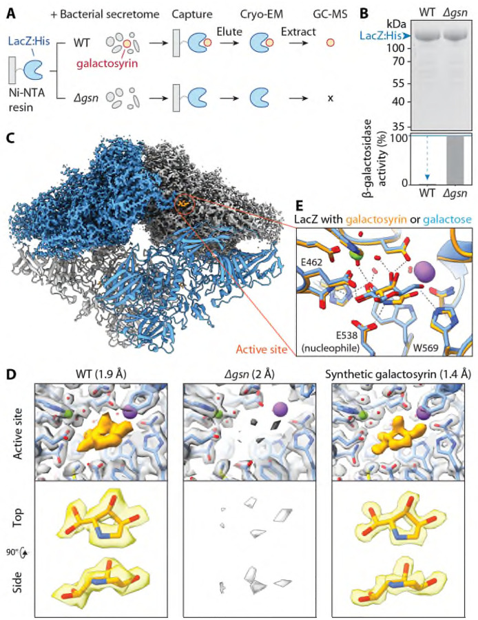
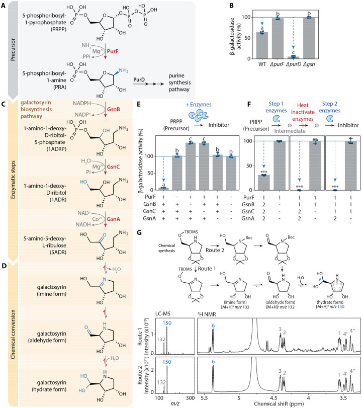
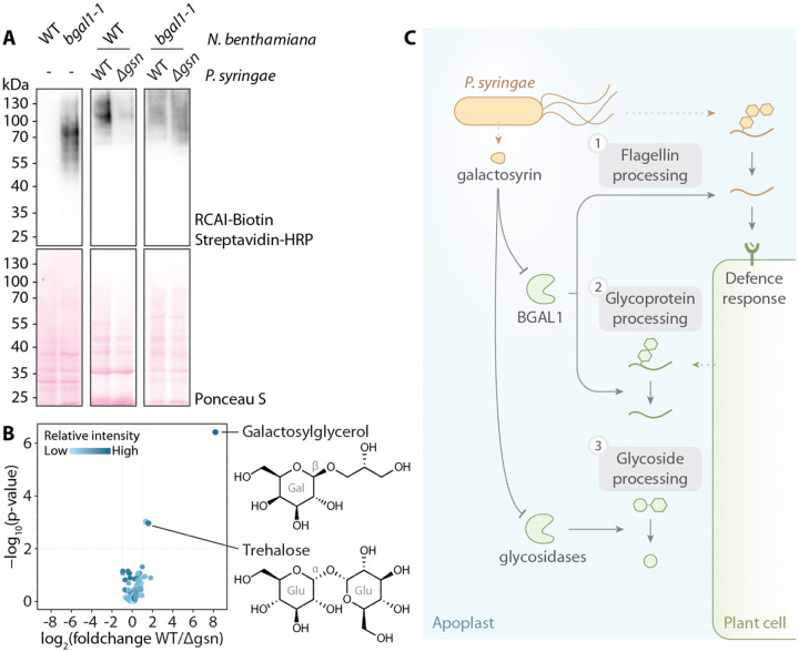
The extracellular space (apoplast) of plants is an important molecular battleground during infection by many pathogens. We previously found that a plant-secreted β-galactosidase BGAL1 acts in immunity by facilitating the release of immunogenic peptides from bacterial flagellin and that Pseudomonas syringae suppresses this enzyme by producing a small molecule inhibitor called galactosyrin. Here, we elucidated the structure and biosynthesis of galactosyrin and uncovered its multifunctional roles during infection. Structural elucidation by cryo-EM and chemical synthesis revealed that galactosyrin is an iminosugar featuring a unique geminal diol attached to the pyrrolidine moiety that mimics galactose binding to the β-galactosidase active site. Galactosyrin biosynthesis branches off from purine biosynthesis and involves three enzymes of which the first is a reductase that is unique in iminosugar biosynthesis. Besides inhibiting BGAL1 to avoid detection, galactosyrin also changes the glycoproteome and metabolome of the apoplast. The manipulation of host glycobiology may be common to plant-associated bacteria that carry putative iminosugar biosynthesis clusters.


Conflict of interest statement

Competing interests: The authors declare no competing interests.

Figures

Fig. 1.
Fig. 1.
Galactosyrin biosynthesis gene cluster and its regulators identified by forward genetics. (A) Genetic screen for galactosyrin-deficient mutants. P. syringae expressing LacZ β-galactosidase was used to create a random transposon insertion mutant library. When plated onto a virulence-inducing medium supplemented with X-gal, galactosyrin-deficient mutants cannot inhibit LacZ, resulting in a darker blue colour. These candidate mutants were validated in an enzymatic assay for the inability to produce galactosyrin. Confirmed mutants were sequenced to identify transposon insertion sites. (B) Histogram with number of transposon insertion sites identified from galactosyrin mutants along the position within the genome, showing four hotspots corresponding to the gsn gene cluster and virulence regulators hrpR, hrpS, hrpL and rhpS. (C) Summary of the roles of genes required for galactosyrin production. The gsn cluster (containing five genes gsnABCDE, PSPTO0834-8) confers galactosyrin biosynthesis. The expression of the gsn cluster is controlled by a regulatory cascade of type III secretion system regulators (RhpS, HrpR, HrpS and HrpL), which controls virulence gene induction during infection. The promotor of the gsn cluster contains the binding site of the HrpL transcriptional activator (hrp box). (D) The gsn cluster confers galactosyrin biosynthesis in P. syringae and E. coli. Bacterial strains were grown in virulence-inducing medium and the supernatant was tested for LacZ inhibition using purified LacZ and substrate FDG (Fluorescein di(-β-D-Galactopyranoside). β-galactosidase activity is reported as a percentage of the activity relative to the mean of the no-inhibitor-control (Δgsn or empty vector). Arrows highlight significant inhibition. Error bars represent standard deviation from 3 replicates. Asterisks indicate statistically significant difference compared to no-inhibitor-control (P < 0.001) using Welch’s t-test. (E) Expression of the gsn cluster is dependent on hrpR, hrpS, hrpL and rhpS. Bacterial strains were grown in virulence-inducing medium, then total RNA was extracted for reverse transcription polymerase chain reaction (RT-PCR) to monitor transcript levels of gsnA, avrPtoB (type III secreted effector gene) and rpoD (reference gene). (F) The gsn cluster is transcribed during infection. Bacteria carrying various promoter:luxCDABE reporter fusion constructs were infiltrated into N. benthamiana leaves and luminescence was imaged at different time points after infection. Signals displayed are scaled to the maximum and minimum within each image. Leaves are outlined with dashed lines. (G) gsn cluster contributes to virulence. Bacterial strains were spray-inoculated on N. benthamiana leaves then bacterial growth (number of bacterial colony forming units (CFU) per cm2 of leaf) was quantified at 3 days post infection. Results from 3 independent experiments with 12 replicates each are plotted in different colours. Asterisks indicate statistically significant difference between strains (P < 0.001) using two-way ANOVA with experiments as blocks.
Fig. 2.
Fig. 2.
Galactosyrin is a hydrated pyrrolidine of a novel iminosugar class. (A) LacZ-galactosyrin complex capture and downstream analyses. A Histidine-tagged β-galactosidase enzyme from E. coli (LacZ:His) immobilised on Ni-NTA beads was used to capture galactosyrin inhibitor from crude bacterial secretome of galactosyrin-producing P. syringae (WT) or the galactosyrin-deficient mutant (Δgsn, negative control). After washing, the complex was eluted and used for cryo-electron microscopy (Cryo-EM), and soluble metabolites were extracted for analysis by gas chromatography-mass spectrometry (GC-MS). (B) Captured LacZ is saturated with galactosyrin. (Top) Total protein stain of eluted samples separated on SDS-PAGE. (Bottom) β-galactosidase activity of each sample measured by FDG assay showing inhibition of WT sample compared to Δgsn. (C) Structure of LacZ-galactosyrin complex from Cryo-EM. The density map is shown for the top half of the structure and a fitted model is shown for the bottom half. Each monomer of LacZ tetramer is coloured differently. (D) Structure of galactosyrin revealed by Cryo-EM. Top: structures of LacZ-galactosyrin complex capture from WT or Δgsn strains and of LacZ incubated with synthetic galactosyrin. Density maps with fitted protein structures show the enzyme active site with the presence and absence of galactosyrin (orange). The resolution of each structure is shown in brackets. Bottom: extracted density map with fitted structure of galactosyrin from top and side view. (E) Galactosyrin mimics galactose binding in the active site. Overlay of structures of the LacZ active site and interacting residues in complex with galactosyrin (orange) or galactose (blue) showing similarity of overall binding pose and positioning of hydroxyl groups. The positive charge on the likely protonated amine nitrogen of galactosyrin can introduce extra electrostatic interaction with the negatively charged catalytic glutamic acid (E538) and cation-pi interaction with the aromatic tryptophan (W569). The stick representation of the molecular structure is coloured by heteroatoms (red:oxygen, blue:nitrogen) while hydrogen is not shown. The green sphere represents Mg2+ and the purple sphere represents Na+. Dashed lines represent hydrogen bonds.
Fig. 3.
Fig. 3.
Galactosyrin biosynthesis branches off from purine biosynthesis by enzymatic and chemical conversion. (A) Galactosyrin biosynthesis branches off from the purine biosynthesis pathway. (B) purF but not purD is required for galactosyrin biosynthesis. Bacterial strains (WT or knockout mutants ΔpurF, ΔpurD, Δgsn) were grown in virulence-inducing MG medium containing purines overnight then the supernatant was tested for inhibitor production. (C) Three gsn-encoded enzymes convert PRA into 5ADR. (D) A plausible 3-step chemical conversion pathway of 5ADR into the final hydrate form of galactosyrin. (E) PurF, GsnB, GsnC and GsnA are required and sufficient for the biosynthesis of galactosyrin from PRPP in vitro. Galactosyrin biosynthesis was reconstructed by mixing PRPP precursor with purified enzymes and their cofactors. For different mixtures, + indicates added enzymes, while - indicates omitted enzymes. (F) PurF, GsnB, GsnC and GsnA act consecutively in galactosyrin biosynthesis. Biosynthesis of galactosyrin was reconstructed by mixing PRPP precursor with purified enzymes and their cofactors in 2 separate steps: [1] enzymes added in the first step to produce an intermediate before heat inactivation of the enzymes, then [2] enzymes added in the second step to complete galactosyrin biosynthesis. (B, E, F) Inhibitor production was tested in an enzyme activity assay with FDG substrate and LacZ enzyme. β-galactosidase activity is reported as a percentage of the activity relative to the mean of no-inhibitor-control (Δgsn for B, all enzymes omitted for E, enzyme 2 omitted for F). Arrows highlight inhibition. Error bars represent standard deviation from 3 replicates. Different letters indicate different groups with statistically significant difference (P < 0.001) using one-way ANOVA and post-hoc Tukey HSD test (for B, E). Asterisks indicate statistically significant difference (P < 0.001) using Welch’s t-test (for F). (G) Both imine and aldehyde forms of galactosyrin spontaneously convert into the hydrate form in water. (Top) Chemical synthesis of galactosyrin using two routes, via imine or aldehyde forms. (Bottom) Products were analysed with liquid chromatography-mass spectrometry (LC-MS) (left) and H1-NMR (right), showing the spectra that correspond to the spontaneously formed hydrate form. Positions within the structure of the hydrate form are numbered and labelled on the corresponding signals in NMR spectra.
Fig. 4.
Fig. 4.
Galactosyrin manipulates multiple aspects of plant apoplast glycobiology. (A) Accumulation of RCAI-positive glycoproteins in the apoplast upon infection is dependent on BGAL1 and galactosyrin production. Proteins were extracted by acetone precipitation of apoplastic fluids from N. benthamiana (wild-type (WT) or BGAL1 knockout mutant (bgal1-1) with or without infection by P. syringae (WT or Δgsn), then separated on SDS-PAGE, blotted and probed with RCAI lectin targeting galactose. (B) Galactosylglycerol and trehalose accumulate in the apoplast during infection dependent on galactosyrin production. Volcano plot of soluble metabolites detected by GC-MS of apoplastic fluids from leaves infected with WT or Δgsn mutant (Table S2). Glucoside components are shown with galactose (Gal), Glucose (Glu) and bond configuration (α or β). (C) P. syringae produces galactosyrin to manipulate multiple aspects of glycobiology inside host plants by inhibiting plant glycosidases in the apoplast. [1] One major target of galactosyrin is the β-galactosidase BGAL1 previously shown to play a role in the processing of glycosylated flagellin to release plant defence elicitor (2). [2] Inhibition of BGAL1 by galactosyrin also interrupts apoplastic glycoprotein processing resulting in the accumulation of galactose-containing glycoproteins. [3] Galactosyrin also disrupts processing of glycosides by glycosidases other than BGAL1, resulting in the accumulation of galactosylglycerol and trehalose in the apoplast.

References

    1. Doehlemann G., Hemetsberger C., Apoplastic immunity and its suppression by filamentous plant pathogens. New Phytol. 198, 1001–1016 (2013). - PubMed
    1. Buscaill P., Chandrasekar B., Sanguankiattichai N., Kourelis J., Kaschani F., Thomas E. L., Morimoto K., Kaiser M., Preston G. M., Ichinose Y., van der Hoorn R. A. L., Glycosidase and glycan polymorphism control hydrolytic release of immunogenic flagellin peptides. Science (80-.). 364 (2019). - PubMed
    1. Wei C., Kvitko B. H., Shimizu R., Crabill E., Alfano J. R., Lin N., Martin G. B., Huang H., Collmer A., A Pseudomonas syringae pv. tomato DC3000 mutant lacking the type III effector HopQ1–1 is able to cause disease in the model plant Nicotiana benthamiana. Plant J. 51, 32–46 (2007). - PubMed
    1. Vencato M., Tian F., Alfano J. R., Buell C. R., Cartinhour S., DeClerck G. A., Guttman D. S., Stavrinides J., Joardar V., Lindeberg M., Bronstein P. A., Mansfield J. W., Myers C. R., Collmer A., Schneider D. J., Bioinformatics-enabled identification of the HrpL regulon and type III secretion system effector proteins of Pseudomonas syringae pv. phaseolicola 1448A. Mol. Plant-Microbe Interact. 19, 1193–1206 (2006). - PubMed
    1. Xie Y., Shao X., Deng X., Regulation of type III secretion system in Pseudomonas syringae. Environ. Microbiol. 21, 4465–4477 (2019). - PubMed

Publication types

LinkOut - more resources