[Preprint]. 2025 Feb 11:2025.02.10.637575.
doi: 10.1101/2025.02.10.637575.

Mammalian oocytes receive maternal-effect RNAs from granulosa cells

Affiliations

Mammalian oocytes receive maternal-effect RNAs from granulosa cells

Caroline A Doherty et al. bioRxiv. .

Abstract

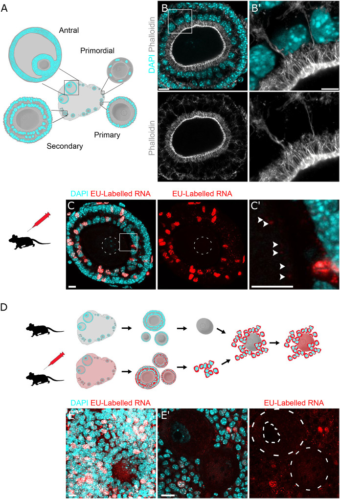
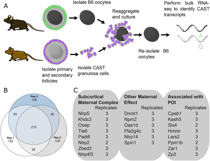
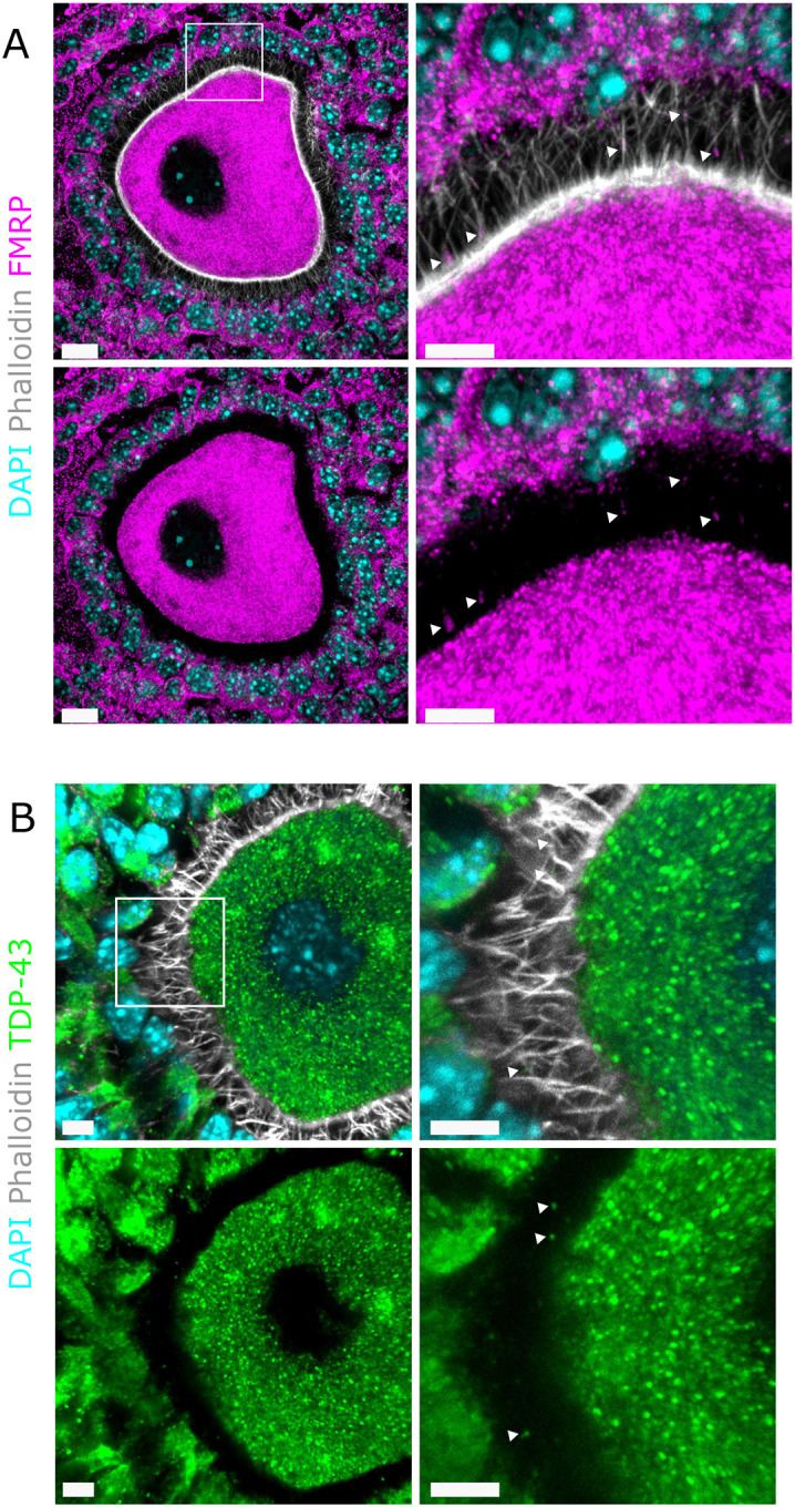
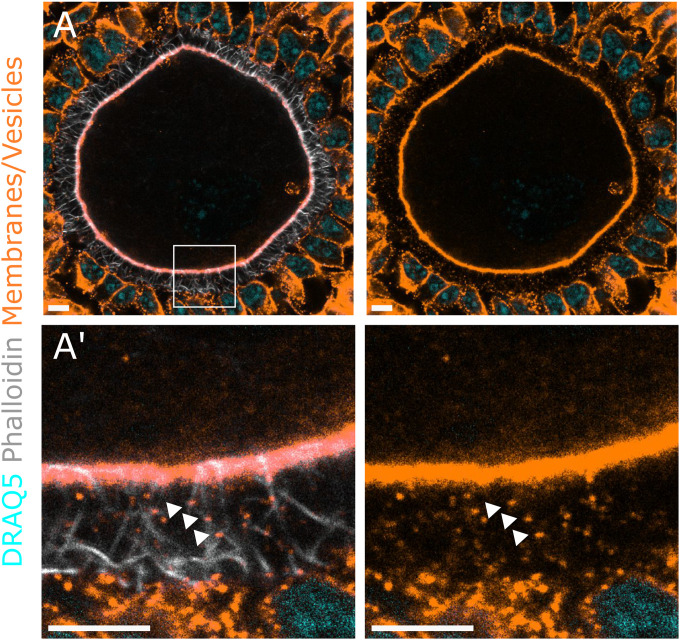
It is currently thought that growing mammalian oocytes receive only small molecules via gap junctions from surrounding support cells, the granulosa cells. From the study of chimeric preantral oocyte and granulosa cell reaggregations, we provide evidence that growing mouse oocytes receive mRNAs from granulosa cells. Among the >1,000 granulosa-transcribed RNAs we identified in the oocyte, those that contribute to proper oocyte maturation and early embryo development were highly enriched. Predicted motifs for two RNA-binding proteins that function in RNA trafficking, FMRP and TDP43, were abundant in the UTRs of the granulosa-derived transcripts. Immunostaining demonstrated that both FMRP and TDP43 co-localize with the actin-rich granulosa cell protrusions that span the zone pellucida and connect to the oocyte, suggesting their role in importing mRNAs. Our results offer the possibility that oocyte failure may not always reflect an intrinsic oocyte deficiency but could arise from insufficient supply of maternal transcripts by granulosa cells during oocyte growth.

Keywords: Fertility; Granulosa cells; Oocyte; RNA transfer; Transzonal projections.


Figures

Figure 1.
Figure 1.. Labeled RNA in granulosa cells can be detected in preantral oocytes
A. Schematic of folliculogenesis: Primordial follicles are quiescent oocytes present from birth that are surrounded by a layer of squamous granulosa cells. Primary follicles are growing follicles that have begun secreting a layer of glycoprotein, the zona pellucida, that separates the oocyte from the granulosa cells. To maintain contact with oocytes across the zona pellucida, granulosa cells grow thin, filopodia-like projections, called transzonal projections (TZPs). Secondary follicles have multiple layers of granulosa cells. Both inner and outer granulosa cells grow TZPs that contact the oocyte. Antral follicles grow a large fluid-filled sac and are the final stage of oogenesis prior to ovulation. B. NSPARC z-projection of an intact mouse multilayered secondary follicle. Granulosa cell nuclei marked with DAPI in cyan and transzonal projections (TZPs) marked with phalloidin in grayscale. A magnification (B’) highlights a TZPs contacting the oocyte from both the inner and outer layers of granulosa cells. Scale bars = 10 um. C. Wholemount confocal z-projection of a secondary follicles processed with Click-iT kit 16 hours after ethynyl uridine IP injection. DNA labelled with DAPI in cyan and nascent RNA labelled with EU in red. Nascent RNA does not appear in the oocyte nucleus (outlined in white dashes) however it does appear in small puncta at the oocyte cortex seen in the magnification (C’) noted with arrowheads. Scale bars = 5 um. D. Re-aggregation scheme whereby unlabeled oocytes are re-aggregated with granulosa cells labeled with EU. The granulosa cells were isolated 16 hours after IP injection of EU. After six days of culture the aggregates are fixed and processed with a Click-iT kit to visualize EU-labeled RNA. E. Wholemount z-projection of reaggregates and single confocal slice (E’). EU-labelled puncta appear in the oocytes, indicating transfer of RNA from granulosa cells to oocytes. Scale bars = 10 um.
Figure 2.
Figure 2.. Identification of granulosa-derived transcripts in preantral oocytes
A. Schematic of follicle re-aaggregation protocol combining primary or secondary stage B6 oocytes (black) with Cast granulosa cells (purple). B. Out of the top 500 genes with CAST SNPs with most abundant reads identified in RNA-seq from B6 oocytes, 273 were present in all three follicle reaggregation replicates (Rep). C. Relevant transcripts identified that are associated with the subcortical maternal complex, maternal-effect genes, and genes associated with primary ovarian insufficiency.
Figure 3.
Figure 3.. RNA-Binding Proteins predicted to bind to trafficked RNAs localize in TZPs
A. Immunostaining and super-resolution microscopy demonstrate co-localization of FMRP (pink) with TZPs (stained with phalloidin in grey; see arrowheads) and “docking” of FMRP with the oocyte cortex. DAPI marks nuclei in turquoise. Scale bars = 10 um. B. TDP-43 (green) similarly localizes with TZPs (grey; see arrowheads). Scale bars = 5 um.
Figure 4.
Figure 4.. Vesicles co-localize with TZPs
A. Wholemount confocal slice and magnification (A’) of a mouse follicle expressing the mTmG reporter, labeling membranes and vesicles with tdTomato in orange. TZPs labeled with phalloidin in grayscale and DNA labelled with DRAQ5 in cyan. Magnified image demonstrates co-localization of vesicles with TZPs. Scale bars = 5 um.

References

    1. Liu K., Rajareddy S., Liu L., Jagarlamudi K., Boman K., Selstam G., and Reddy P. (2006). Control of mammalian oocyte growth and early follicular development by the oocyte PI3 kinase pathway: New roles for an old timer. Dev Biol 299, 1–11. 10.1016/J.YDBIO.2006.07.038. - DOI - PubMed
    1. Yurttas P., Morency E., and Coonrod S.A. (2010). Use of proteomics to identify highly abundant maternal factors that drive the egg-to-embryo transition. Reproduction 139, 809–823. 10.1530/REP-09-0538. - DOI - PubMed
    1. Mitchell L.E. (2022). Maternal effect genes: Update and review of evidence for a link with birth defects. Human Genetics and Genomics Advances 3, 100067. 10.1016/J.XHGG.2021.100067. - DOI - PMC - PubMed
    1. Li L., Zheng P., and Dean J. (2010). Maternal control of early mouse development. Development 137, 859–870. 10.1242/DEV.039487. - DOI - PMC - PubMed
    1. Doherty C.A., Amargant F., Shvartsman S.Y., Duncan F.E., and Gavis E.R. (2021). Bidirectional communication in oogenesis: a dynamic conversation in mice and Drosophila. Trends Cell Biol, 1–13. 10.1016/j.tcb.2021.11.005. - DOI - PMC - PubMed

Publication types

LinkOut - more resources