[Preprint]. 2025 Feb 15:2025.02.11.637683.
doi: 10.1101/2025.02.11.637683.

Genomic and functional adaptations in guanylate-binding protein 5 (GBP5) highlight specificities of bat antiviral innate immunity

Affiliations

Genomic and functional adaptations in guanylate-binding protein 5 (GBP5) highlight specificities of bat antiviral innate immunity

Amandine Le Corf et al. bioRxiv. .

Abstract

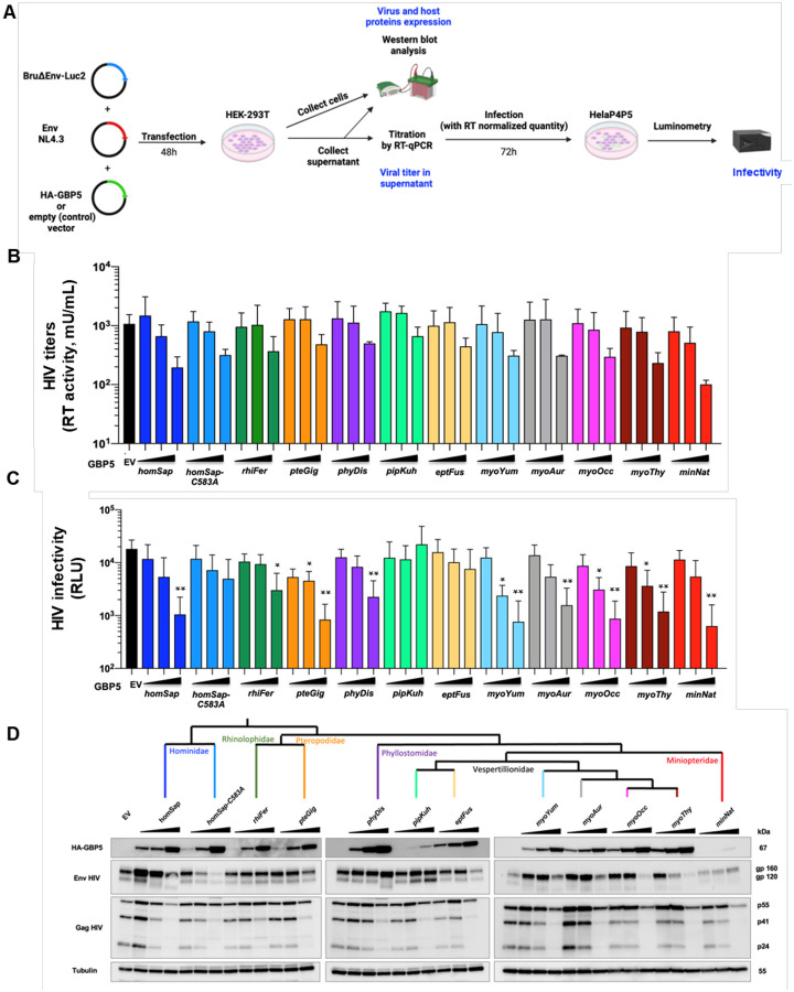
Bats are asymptomatic reservoirs of several zoonotic viruses. This may result from long-term coevolution between viruses and bats, that have led to host adaptations contributing to an effective balance between strong antiviral responses with innate immune tolerance. To better understand these virus-host interactions, we combined comparative transcriptomics, phylogenomics and functional assays to characterize the evolution of bat innate immune antiviral factors. First, we stimulated the type I interferon immune pathway in Myotis yumanensis primary cells and identified guanylate-binding protein 5 (GBP5) as the most differentially expressed interferon-stimulated gene (ISG). Phylogenomic analyses showed that bat GBP5 has been under strong episodic positive selection, with numerous rapidly evolving sites and species-specific gene duplications, suggesting past evolutionary arms races. Functional tests on GBP5 orthologs from ten bat species covering the >60 million years of Chiroptera evolution revealed species- and virus-specific restrictions against RNA viruses (retrovirus HIV, and rhabdoviruses European bat lyssavirus and VSV), which are typical signatures of adaptations to past viral epidemics. Interestingly, we also observed a lineage-specific loss of the GBP5 prenylation motif in the common ancestor of Pipistrellus and Eptesicus bats, associated with different GBP5 subcellular localization and loss of antiviral functions. Resurrection of the ancestral prenylation motif in Eptesicus fuscus GBP5 rescued its subcellular localization, but not the complete antiviral activities, suggesting that additional determinants are necessary for the antiviral restriction. Altogether, our results highlight adaptations that contribute to bat specific immunity and provide insights into the functional evolution of antiviral effector GBP5.

Keywords: Chiroptera; EBLV-1; GBP5; Guanylate-binding proteins; HIV; Rhabdovirus; antiviral factor; bats; innate immunity; molecular arms-race; positive selection; retrovirus; virus-host coevolution.


Figures

Figure 1.
Figure 1.. GBP5 is the most upregulated ISG in Myotis yumanensis primary cells upon interferon immune stimulation.
A, Photo of a Myotis yumanensis bat at a sampling site (Credit: Elise Lauterbur). B, Primary cell line derived from biopsy wing punches. C, Schematic of the workflow from bat sampling to RNA sequencing analyses. Bat primary cell lines were similarly derived from three M. yumanensis individuals and treated with, or without, universal type I IFN to trigger ISG expression. Total RNA was extracted and sequenced, followed by analyses to identify differentially expressed genes between stimulated cells compared to control cells. D, Volcano plot representing the differential gene expression between untreated and IFN-stimulated cells. Genes significantly (p<0.05) differentially expressed (DE) are colored in red. Several known ISGs are highlighted on the graphic. GBP5 is the most upregulated ISG. E, Comparative analysis of differential gene expression (Log2FC, Log 2 Fold Change) upon type I IFN treatment of GBP5 in M. yumanensis cells with six mammalian species cells in similar experimental setup (; details in Methods).
Figure 2.
Figure 2.. Mammalian GBP5 has evolved under episodic positive selection, with strong diversification in rodents and bats.
A, Phylogenetic analysis of GBP5 across mammals. The phylogeny was built from a PRANK codon alignment of 348 GBP5 homologous sequences. Maximum likelihood phylogenetic tree was built with IQ-TREE with 1000-bootstrap replicates for statistical support (see Data availability). Branches under significant positive selection (p-value <0.05) assigned by aBSREL are thickened and in red. The scale bar indicates the number of substitutions per site. B, Evidence of positive selection in GBP5 from several mammalian orders. Positive selection analyses were performed with BUSTED using a PRANK codon alignment for each mammalian order. OWM, Old world monkeys. Species silhouettes are from https://www.phylopic.org.
Figure 3.
Figure 3.. GBP5 has evolved under strong positive selection in bats.
A, Phylogenetic and positive selection analyses of bat GBP5. Maximum likelihood phylogenetic tree of bat GBP5 was built with IQ-TREE and statistical support is from 1000 bootstrap replicates (values are shown below branches). Branches under significant positive selection (p-value <0.05) assigned by aBSREL are in red and the corresponding estimated values of ω are reported in panel B. The scale bar indicates the number of substitutions per site. The black arrows identify species with GBP5 genes that were functionally tested in this study. 1.1 and 1.2 identify GBP5 duplicates within a given bat species. B, Evidence of lineage-specific positive selection during bat GBP5 evolution. aBSREL identified at least six branches under significant positive selection. ω1 and ω2, estimation of ω in the rate class not allowing positive selection, and allowing positive selection (ω>1), with % of sites in this class in parentheses, respectively. C, Sites under positive selection (PS) in bat GBP5. Site-specific positive selection analyses were performed using FUBAR, MEME and FEL from HYPHY/Datamonkey,. Only the sites above the indicated “statistically significant cut-off” are shown (PP, posterior probabilities for FUBAR; p-value for MEME and FEL). In bold are the sites identified by several methods. Nb of PSS, number of positively selected sites. “PS sites” numbering is according to the codon numbering in the PRANK codon alignment. D, Schematic representation of GBP5 with its functional domains and the herein identified sites under positive selection (red arrows at the top). LG domain, large GTPase domain. MD, Middle domain. GED, GTPase effector domain. The size of the domains is not to scale. Residues identified by at least two positive selection methods are highlighted in bold. In blue, sites or motifs involved in known functions in human GBP5.
Figure 4.
Figure 4.. Natural variation in subcellular localization of bat GBP5s.
TZM-bl cells were transfected with a plasmid coding for indicated HA-GBP5 species proteins: 10 bat orthologs, and 2 human GBP5s: wt and mutant C583A. Two days post-transfection, GBP5 localization was analyzed by confocal fluorescence microscopy with the indicated marker. Nuclei and trans-golgi-network (TGN) were stained with DAPI and anti-TGN46, respectively. A, All the channels and a zoom are shown for Homo sapiens, the mutant Homo sapiens-C583A, Myotis yumanensis and Eptesicus fuscus. B, Only the merge is shown for the remaining bat species. The complete panel is shown in Fig. S4. The pictures present representative results observed in 3 independent experiments. Scale bar indicates 15 μm.
Figure 5.
Figure 5.. Species-specificity in bat GBP5 restriction of retrovirus (HIV-1).
A, Experimental setup. Briefly, HEK-293T cells were transfected with plasmids coding for HA-GBP5 or the empty vector (control), and for HIV-1 LAI genome and Luciferase reporter (Bru∂EnvLuc2 vector), NL4.3 Envelope and Rev. 48h post-transfection, cells were harvested for western-blot analysis, and supernatant for western-blot analysis after ultracentrifugation, titration of virus by RT-qPCR (RT activity), and infection of the RT-normalized viruses in HelaP4P5 cells. Infection was quantified 72h later by luminescence from the viral-encoded Luciferase reporter. B, HIV-1 titers in the supernatants as quantified by RT activity (mU/ml) in the context of a dose of HA-GBP5 (1, 2 or 4 μg) or control vector (EV). The corresponding species of GBP5 is shown (name follows the UCSC nomenclature, three letters from genus followed by three letters from species). C, Corresponding HIV-1 infectivity (RLU, Relative light units). Statistics versus the corresponding control condition: *, p value <0.05, **, p value <0.01. D, Corresponding western blot analysis of HA-GBP5, HIV-1 Env and HIV-1 Gag, and Tubulin (loading control) from the lysates of the HIV-1 producer cells in the context of a GBP5 dose. The cladogram at the top shows the phylogenetic relationships of the tested species. The western blot of the purified virion fraction (supernatant) is in Fig. S5.
Figure 6.
Figure 6.. Bat GBP5 restriction is both species- and virus-specific.
A, Experimental setup similar to Fig. 5A but with viral pseudotyping with VSVg (VSV condition) or EBLV-1 envelope (EBLV-1 condition). B, Infectivity of RT-normalized pseudotyped-viruses produced in the presence of GBP5, normalized to the condition without GBP5 (EV, Empty vector control) at 100%. The cladogram on the left shows the phylogenetic relationships of the tested GBP5 species. RLU, Relative light units. Statistics versus the corresponding control condition: *, p value <0.05, **, p value <0.01, ***, p value <0,001.
Figure 7.
Figure 7.. Ancestral reconstruction of the prenylation motif in Eptesicus fuscus relocalizes GBP5 to the TGN, but is not sufficient to retrieve full antiviral functions.
A, Ancestral state sequence reconstruction upstream of the Eptesicus fuscus-CaaX prenylation motif. C-terminal end of the protein alignment of the 10 bat GBP5s tested in functional assays (asterisk, stop codon). Phylogenetic tree was used to infer the ancestral sequence of the C terminal region, the branch where the prenylation motif was lost by a premature stop codon is annotated on the tree. The site of mutagenesis for reconstruction is indicated by the blue arrow. B, Reconstruction of the C-ter relocalizes Eptesicus fuscus GBP5-CaaX to the trans-golgi network (TGN). Briefly, TZM-bl cells were transfected with plasmids encoding HA-GBP5s and, 48h later, were analyzed by confocal fluorescence microscopy. Nuclei and TGN were stained with DAPI and anti-TGN46, respectively. Scale bar indicates 15 μm. C, GBP5 mean intensity at the Golgi versus the total cell was quantified for the wild-type eptFus and the mutant eptFus-CaaX. Each dot corresponds to one cell. Replicates are grouped according to dot color. D, Pearson coefficient correlation per cell calculated between GBP5 and TGN signals for the wild-type eptFus and the mutant eptFus-CaaX. E-G, Ancestral reconstruction of the prenylation CaaX did not increase Eptesicus fuscus GBP5 antiviral functions against infectivity. E, Infectivity of RT-normalized HIV-1 pseudotyped viruses in the presence of GBP5, normalized to the condition without GBP5 (EV, Empty vector control) at 100%. Dose of GBP5 plasmids: 1, 2, 4 μg. Experimental setup as in Fig. 5A. RLU, Relative light units. Viral titers are shown in Fig. S5. F, Corresponding western-blot showing the expression of HA-GBP5, HIV-1 Env and Gag in the viral producer cells with beta-actin as loading control (kDa, on the right). G, Infectivity of RT-normalized VSVg or EBLV-1g pseudotyped viruses in the presence of GBP5 variants, normalized to the condition without GBP5 (empty vector control) at 100%. Experimental setup as in Fig 6A. H, 3D protein structure prediction (AlphaFold) of the reconstructed Eptesicus fuscus-CaaX GBP5 dimer. Colored and grey chains each correspond to a monomer. Blue, GTPase domain. Green, hinge domain. Yellow, middle domain. Orange, catalytic domain. Red, residues different from Myotis yumanensis. Statistics versus the corresponding control condition: **, p-value < 0.01.

References

    1. Tenthorey J. L., Emerman M. & Malik H. S. Evolutionary Landscapes of Host-Virus Arms Races. Annu. Rev. Immunol. 40, 271–294 (2022). - PubMed
    1. Duggal N. K. & Emerman M. Evolutionary conflicts between viruses and restriction factors shape immunity. Nat. Rev. Immunol. 12, 687–695 (2012). - PMC - PubMed
    1. Irving A. T., Ahn M., Goh G., Anderson D. E. & Wang L.-F. Lessons from the host defences of bats, a unique viral reservoir. Nature 589, 363–370 (2021). - PubMed
    1. Ahn M. et al. Dampened NLRP3-mediated inflammation in bats and implications for a special viral reservoir host. Nat. Microbiol. 4, 789–799 (2019). - PMC - PubMed
    1. Ahn M., Cui J., Irving A. T. & Wang L.-F. Unique Loss of the PYHIN Gene Family in Bats Amongst Mammals: Implications for Inflammasome Sensing. Sci. Rep. 6, 21722 (2016). - PMC - PubMed

Publication types

LinkOut - more resources