[Preprint]. 2025 Feb 13:2025.02.12.637895.
doi: 10.1101/2025.02.12.637895.

A connection between Vibrio cholerae motility and inter-animal transmission

Affiliations

A connection between Vibrio cholerae motility and inter-animal transmission

Ian W Campbell et al. bioRxiv. .

Update in

Abstract

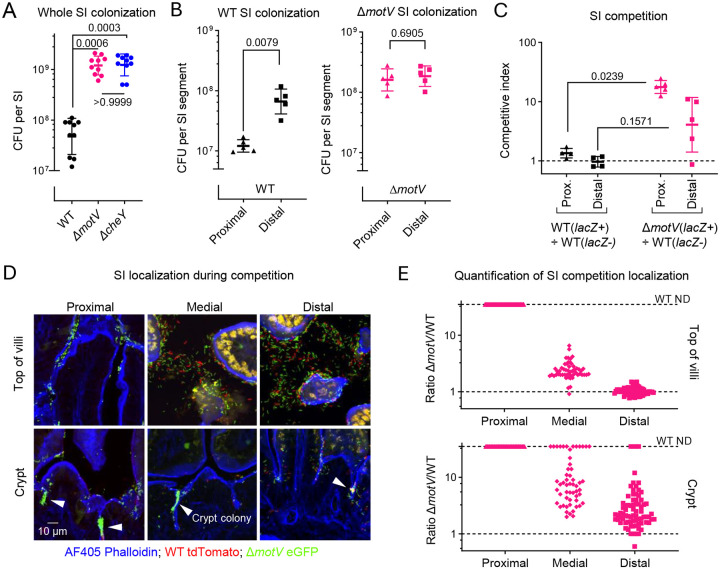
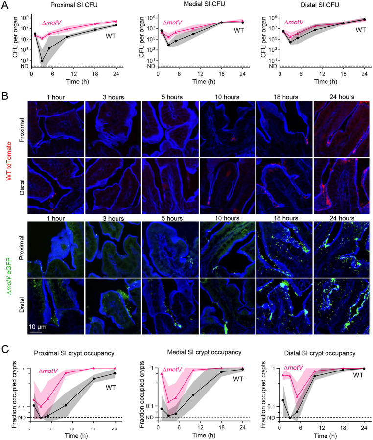
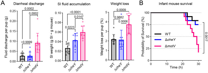
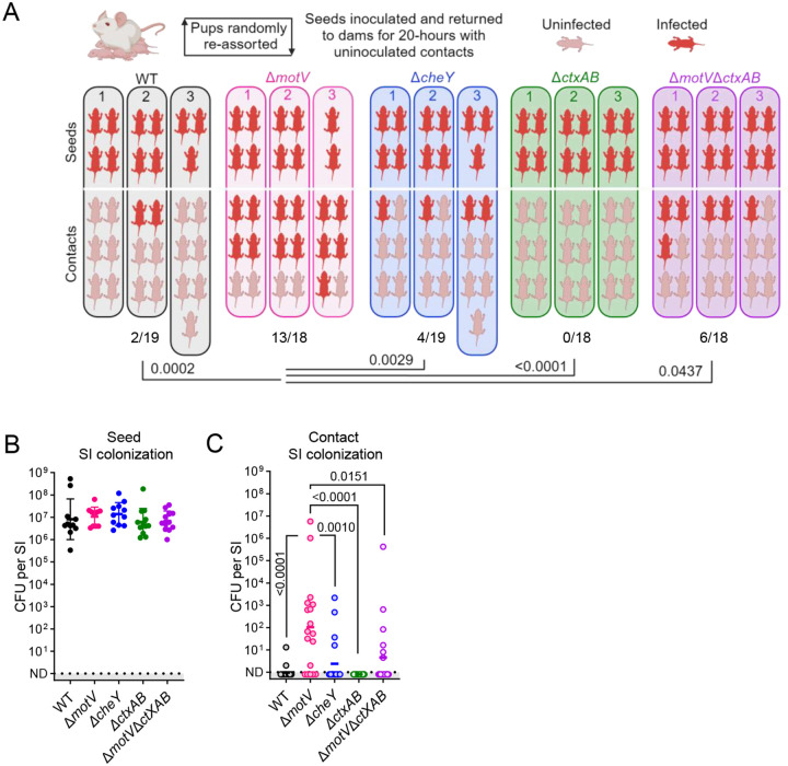
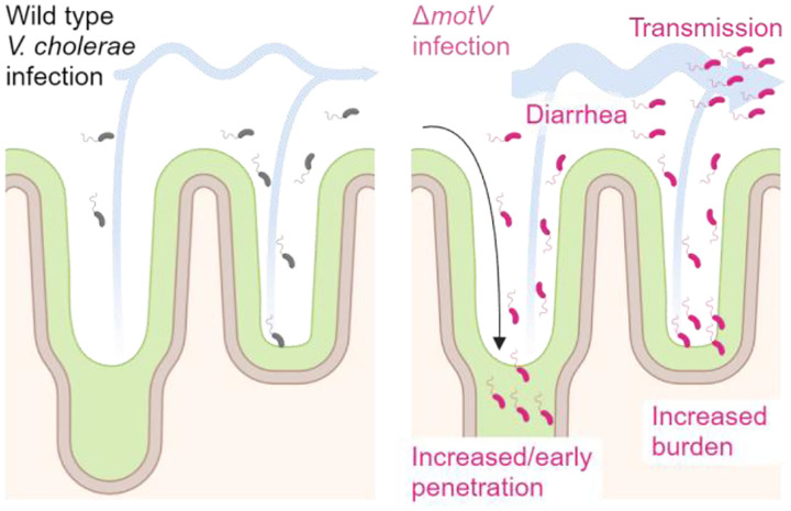
Outbreaks of cholera are caused by the highly transmissive pathogen Vibrio cholerae. Here, a transposon screen revealed that inactivation of the V. cholerae motility-linked gene motV increases infant mouse intestinal colonization. Compared to wild-type V. cholerae, a ΔmotV mutant, which exhibits heightened motility in the form of constitutive straight swimming, localizes to the crypts earlier in infection and over a larger area of the small intestine. Aberrant localization of the mutant was associated with an increased number of V. cholerae initiating infection, and elevated pathogen burden, diarrhea, and lethality. Moreover, the deletion of motV causes V. cholerae to transmit from infected suckling mice to naïve littermates more efficiently. Even in the absence of cholera toxin, the ΔmotV mutant continues to transmit between animals, although less than in the presence of toxin, indicating that phenotypes other than cholera toxin-driven diarrhea contribute to transmission. Collectively, this work provides experimental evidence linking intra-animal bottlenecks, colonization, and disease to inter-animal transmission.


Conflict of interest statement

Competing Interests The authors declare no competing interests.

Figures

Figure 1 |
Figure 1 |. Tn-seq screen reveals that disruption of motV increased V. cholerae infant mouse colonization.
10 CD1 pups were intragastrically inoculated with ~5×10⁷ CFU of a mariner transposon library. 18-hours later, the SIs were homogenized and pooled, and after overnight growth on LB plates, transposon insertion sites were determined by sequencing (pooled litter). For comparison, the same library was passaged overnight on LB plates (LB library). A, Volcano plot comparing the fold-change in gene insertion frequency between the pooled litter and LB library. Colonization factors are genes with P value <0.01 and log2 fold-change <−2. Data for the tcp operon (toxin-coregulated pilus) and flagellar assembly genes (categorized from KEGG database vch02040) are expanded in B. C, Transposon insertion frequency across the genome highlighting increased insertion frequency in cheR, cheY, and motV from the pooled intestinal samples.
Figure 2 |
Figure 2 |. Deletion of motV increased V. cholerae SI colonization, especially in the proximal SI and within the crypts.
CD1 pups were intragastrically inoculated with ~3 × 10⁶ CFU of the indicated V. cholerae strains, and 18-hours later the whole intestine (A) or the indicated SI segments (B) were plated on selective media to determine V. cholerae colony-forming units (CFU). C, CD1 pups were infected with ~2 × 10⁶ CFU of a ~1:1 ratio of lacZ+ to lacZ V. cholerae, and the competitive index was calculated as the ratio of lacZ+ to lacZ CFU in the indicated SI segment 18-hours after inoculation divided by the ratio of lacZ+ to lacZ CFU in the inoculum. A and C, Kruskal-Wallis test with Dunn’s multiple comparison correction. B, Mann-Whitney test. A-C, Geometric mean and standard deviation. D, Images of 10 μm cryosections of the indicated SI sections 18-hours after inoculation with ~2 × 10⁶ CFU of a ~1:1 ratio of WT lacZ::tdTomato V. cholerae (red) and ΔmotV lacZ::eGFP V. cholerae (green). Sections were stained with AF405-conjugated phalloidin (blue) and imaged on a spinning disk confocal microscope. Arrowheads show V. cholerae microcolonies. E, Quantification of the ratio of ΔmotV:WT V. cholerae cells at the top(s) of the villi and in the crypts of the SI. When WT was not detected (ND), the ratio was given a value of 35 as an upper limit. V. cholerae were differentiated from background fluorescence by comma/rod morphology.
Figure 3 |
Figure 3 |. The ΔmotV mutant localizes in the SI crypts earlier during infection than WT V. cholerae.
CD1 pups were intragastrically inoculated with ~2 × 10⁶ CFU of either WT lacZ::tdTomato V. cholerae (red) or ΔmotV lacZ::eGFP V. cholerae (green). CFU (A) and cryosections were collected over 24-hours from the indicated SI sections. Sections were stained with AF405-conjugated phalloidin (blue) and imaged on a spinning disk confocal microscope (B). C, Quantification of the number of crypts per field occupied by at least one V. cholerae in the proximal, medial, and distal SI. ND, V. cholerae not detected in any field. 4 animals per timepoint per strain. A, C, Geometric mean and standard deviation.
Figure 4 |
Figure 4 |. V. cholerae lacking motV evaded the colonization bottleneck.
A, Diagram of bacterial population dynamics during infection. The dose of the inoculum and the burden of the pathogen in tissue is enumerated by plating for CFU on selective media. The founding population is the number of unique cells from which the observed population originated and is measured by the loss of barcode diversity (represented as colors) between the inoculum and the observed population. Founders are calculated by STAMPR and expressed as Nr. The bottleneck is the loss of cells between the dose and founders, and net expansion is the gain between founders and burden. B, Dose, founders, and burden of WT and ΔmotV V. cholerae in the SI of CD1 pups 18-hours post-inoculation. WT, 5 pups; ΔmotV, 10 pups. Mann-Whitney test. Geometric mean and standard deviation.
Figure 5 |
Figure 5 |. Deletion of motV increased V. cholerae diarrheaogenicity.
A, CD1 pups were intragastrically inoculated with the indicated V. cholerae strains and individually housed for 18-hours. The bedding was weighed prior to and after infection to determine fluid discharge. Fluid accumulation in the SI 18-hours post inoculation was measured by comparing the weight of the animal to the weight of the SI. Weight loss was determined by comparing animal weight before and after infection. Mean and standard deviation. One-way ANOVA with Turkey’s multiple comparison correction. B, CD1 pups were intragastrically inoculated with the indicated V. cholerae strains, returned to dams for care, and monitored until the predetermined 30-hour endpoint. Survival kinetics with Mantel-Cox test.
Figure 6 |
Figure 6 |. Deletion of motV increased inter-animal transmission.
Transmission assays were performed by randomly reassorting CD1 pups, challenging ~1/3 of the litter with the indicated strain of V. cholerae (seeds), and returning them to foster-dams with naïve littermates (contacts). 20-hours later, transmission was determined by enumerating CFU in the SI. Experimental groups: WT, ΔmotV, and ΔcheY; ΔctxAB and ΔmotV ΔctxAB. A, Experimental results divided by litter. Uninfected contacts, 0 CFU detected; infected, ≥1 CFU identified. B, Seed SI CFU determined 20-hours post inoculation. Geometric mean and standard deviation. C, Contact SI colonization determined after 20-hours of cohousing with seed animals. Geometric mean. Kruskal-Wallis test with Dunn’s multiple comparison correction.
Figure 7 |
Figure 7 |. Model connecting the heightened inter-animal transmission of ΔmotV V. cholerae to aberrant motility, bottleneck evasion, increased burden, and increased diarrheaogenicity.
We propose that the ΔmotV mutant’s constitutive and rapid motility enables V. cholerae to penetrate deeper into the more proximal crypts of the SI early during infection and that this aberrant pattern of localization accounts for its hyper-transmissibility. The expansion of the V. cholerae intestinal niche permits more of the inoculum to survive the colonization bottleneck and replicate, increasing the burden of V. cholerae in the intestine and leading to greater diarrhea. Greater burden and diarrheal flux facilitate the excretion of the pathogen into the environment, promoting transmission.

References

    1. Clemens J. D., Nair G. B., Ahmed T., Qadri F. & Holmgren J. Cholera. The Lancet 390, 1539–1549 (2017). - PubMed
    1. Sugimoto J. D. et al. Household Transmission of Vibrio cholerae in Bangladesh. PLoS Negl Trop Dis 8, e3314 (2014). - PMC - PubMed
    1. Eisenberg M., Robertson S. & Tien J. Identifiability and estimation of multiple transmission pathways in cholera and waterborne disease. J Theor Biol 324, (2013). - PubMed
    1. Domman D. et al. Defining endemic cholera at three levels of spatiotemporal resolution within Bangladesh. Nature Genetics 2018 50:7 50, 951–955 (2018). - PMC - PubMed
    1. Herrington D. A. et al. Toxin, toxin-coregulated pili, and the toxR regulon are essential for Vibrio cholerae pathogenesis in humans. Journal of Experimental Medicine 168, 1487–1492 (1988). - PMC - PubMed

Publication types

LinkOut - more resources