. 2025 Feb 18:17:187-200.
doi: 10.2147/BCTT.S488200. eCollection 2025.

Radiomics Integration of Mammography and DCE-MRI for Predicting Molecular Subtypes in Breast Cancer Patients

Affiliations

Radiomics Integration of Mammography and DCE-MRI for Predicting Molecular Subtypes in Breast Cancer Patients

Xianwei Yang et al. Breast Cancer (Dove Med Press). .

Abstract

Background: Accurate identification of the molecular subtypes of breast cancer is essential for effective treatment selection and prognosis prediction.

Aim: This study aimed to evaluate the diagnostic performance of a radiomics model, which integrates breast mammography and dynamic contrast-enhanced magnetic resonance imaging (DCE-MRI) in predicting the molecular subtypes of breast cancer.

Methods: We retrospectively included 462 female patients with pathologically confirmed breast cancer, including 53 cases of triple-negative, 94 cases of HER2 overexpression, 95 cases of luminal A, and 215 cases of luminal B breast cancer. Radiomics analysis was performed using FAE software, wherein the radiomic features were examined about the hormone receptor status. The performance of the model was evaluated using the area under the receiver operating characteristic curve (AUC) and accuracy.

Results: In multivariate analysis, radiomic features were the only independent predictive factors for molecular subtypes. The model that incorporates multimodal fusion features from breast mammography and DCE-MRI images exhibited superior overall performance compared to using either modality independently. The AUC values (or accuracies) for six pairings were as follows: 0.648 (0.627) for luminal A vs luminal B, 0.819 (0.793) for luminal A vs HER2 overexpression, 0.725 (0.696) for luminal A vs triple-negative subtype, 0.644 (0.560) for luminal B vs HER2 overexpression, 0.625 (0.636) for luminal B vs triple-negative subtype, and 0.598 (0.500) for triple-negative subtype vs HER2 overexpression.

Conclusion: The radionics model utilizing multimodal fusion features from breast mammography combined with DCE-MRI images showed high performance in distinguishing molecular subtypes of breast cancer. It is of significance to accurately predict prognosis and determine treatment strategy of breast cancer by molecular classification.

Keywords: breast cancer; magnetic resonance; mammography; molecular subtypes; radiomics.


Conflict of interest statement

The authors declare no competing interests in this work.

Figures

Figure 1
Figure 1
A 45-year-old female patient was diagnosed with infiltrating ductal carcinoma of the breast. (A-B) Axial images display irregular shapes, spiculated margins, and heterogeneous enhancement of the mass (indicated by arrows). (C) The gradual fusion of the volume of interest into the contour image. (D and E) left medial oblique view. (D) The white arrow shows an irregular mass in the upper quadrant of the left breast with irregular shape and marginal burrs; (E) the white arrow shows an lesion area; (F and G) left nipple-tail view. (F) The white arrow shows the lesion shape is irregular, with visible lobes and burrs. (G) The white arrow shows the lesion area of interest.
Figure 2
Figure 2
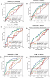
ROC curve for the mammography radiomics model. (A) The AUC of the model constructed based on luminal A and luminal B reaches 0.589 and 0.550 in the verification and test sets, respectively. (B) The AUC of the model constructed based on Luminal A and Her2 overexpression reached 0.622 and 0.612 in the validation and test sets, respectively. (C) The AUC of the models built based on luminal A and TNBC reached 0.506 and 0.608 on the verification and test sets, respectively. (D) The AUC of the models built based on luminal B and HER2 reached 0.592 and 0.563 on the verification and test sets, respectively. (E) The AUC of the models built based on luminal B and TNBC reached 0.557 and 0.602 on the validation and test sets, respectively. (F) The AUC of the models built based on TNBC and HER2 reached 0.545 and 0.571 on the verification and test sets, respectively. HER2+, Human Epidermal Growth Factor Receptor 2+. TNBC, Triple-Negative Breast Cancer.
Figure 3
Figure 3
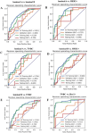
ROC curve for the DCE-MRI radiomics model. (A) The AUC of the model constructed based on luminal A and luminal B reached 0.603 and 0.709 in the verification and test sets, respectively. (B) The AUC of the model constructed based on Luminal A and HER2+ overexpression reached 0.721 and 0.690 in the verification and test sets, respectively. (C) The AUC of the model constructed based on luminal A and TNBC reached 0.681 and 0.600 in the validation set and test set, respectively. (D) The AUC of the model constructed based on luminal B and HER2+ reached 0.602 and 0.601 in the verification and test sets, respectively. (E) The AUC of the model constructed based on luminal B and TNBC reached 0.537 and 0.559 in the verification and test sets, respectively. (F) The AUC of the models built based on TNBC and HER2 reached 0.723 and 0.554 in the verification set and test set, respectively. HER2+, Human Epidermal Growth Factor Receptor 2+. TNBC, Triple-Negative Breast Cancer.
Figure 4
Figure 4
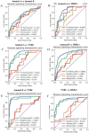
ROC curve for the combined mammography and DCE-MRI radiomics model. (A) The AUC of the models built based on luminal A and luminal B reached 0.615 and 0.698 on the verification and test sets, respectively. (B) The AUC of the models constructed based on luminal A and HER2+ overexpression reached 0.645 and 0.819 in the validation and test sets, respectively. (C) The AUC of the models built based on luminal A and TNBC reached 0.693 and 0.667 on the validation set and test set, respectively. (D) The models built based on luminal B and HER2+ achieved an AUC of 0.682 and 0.644 on the verification and test sets, respectively. (E) The AUC of the models built based on luminal B and TNBC reached 0.626 and 0.625 on the validation set and test set, respectively. (F) The AUC of the models built based on TNBC and HER2+ reached 0.687 and 0.589 on the verification set and test set, respectively. HER2+, Human Epidermal Growth Factor Receptor 2+. TNBC, Triple-Negative Breast Cancer.

References

    1. Bray F, Ferlay J, Soerjomataram I, et al. Global cancer statistics 2018: GLOBOCAN estimates of incidence and mortality worldwide for 36 cancers in 185 countries. CA. 2018;68:394–424. doi:10.3322/caac.21492 - DOI - PubMed
    1. Sung H, Ferlay J, Siegel RL, et al. Global cancer statistics 2020: GLOBOCAN estimates of incidence and mortality worldwide for 36 cancers in 185 countries. CA. 2021;71:209–249. doi:10.3322/caac.21660 - DOI - PubMed
    1. Perou CM, Sørlie T, Eisen MB, et al. Molecular portraits of human breast tumours. Nature. 2000;406:747–752. doi:10.1038/35021093 - DOI - PubMed
    1. Cho N. Imaging features of breast cancer molecular subtypes: state of the art. J Pathol Transl Med. 2021;55:16–25. doi:10.4132/jptm.2020.09.03 - DOI - PMC - PubMed
    1. Bedard PL, Hansen AR, Ratain MJ, et al. Tumour heterogeneity in the clinic. Nature. 2013;501(7467):355–364. doi:10.1038/nature12627 - DOI - PMC - PubMed

LinkOut - more resources