Multicenter Study
. 2025 Feb 5;27(2):euaf003.
doi: 10.1093/europace/euaf003.

Evoked delayed potential ablation for post-myocardial infarction ventricular tachycardia: results from a large prospective multicentre study

Affiliations
Multicenter Study

Evoked delayed potential ablation for post-myocardial infarction ventricular tachycardia: results from a large prospective multicentre study

Marta de Riva et al. Europace. .

Abstract

Aims: The optimal substrate ablation approach for post-myocardial infarction (MI) ventricular tachycardia (VT) is unknown. Proposed ablation targets are prone to individual interpretation making the ablation outcome potentially operator dependent. Evoked delayed potentials (EDPs) are a well-defined target. Evoked delayed potential ablation was effective in preventing post-MI VT recurrence in a prior study. The aims of this study were to assess long-term outcomes of EDP ablation in a large multicentre cohort of post-MI patients and to compare ablation outcomes between centres with and without prior experience in EDP ablation.

Methods and results: Patients with post-MI VT undergoing ablation in one centre performing EDP ablation since 2013 and five centres without prior experience in EDP ablation were prospectively included. A uniform mapping protocol including right ventricular extra-stimulation aiming to EDP identification was followed. Ablation endpoints were EDP elimination and VT non-inducibility. Patients were followed for VT recurrence, mortality, heart transplant, and left ventricular assist device implantation. In total, 130 patients were included. The protocol was successfully performed in 99%, and in 94%, EDPs were identified and ablated. In total, 78% of patients were rendered non-inducible. Ventricular tachycardia-free survival was 78% [95% confidence interval (CI) 71-85] and 71% (95% CI 63-80) at 6 and 12 months, respectively. No difference in VT-free survival was observed among centres with and without prior experience in EPD ablation.

Conclusion: In a large multicentre prospective cohort of patients with post-MI VT, EDP ablation resulted in good long-term outcomes. Importantly, VT recurrence rates did not differ among centres with and without prior experience in EDP ablation, indicating that this approach can be easily reproduced by operators previously not familiar with the technique.

Keywords: Evoked delayed potentials; Functional substrate mapping; Myocardial infarction; Substrate ablation; Substrate modification; Ventricular tachycardia.


Conflict of interest statement

Conflict of interest: The Department of Cardiology Leiden received unrestricted research and fellowship grants from Edward Lifesciences, Boston Scientific, Medtronic, Biotronik, and Biosense Webster. A.O. received consulting fees from Abbott, Biosense Webster, Boston Scientific, and Medtronic. K.V. received research grants from Medtronic, Biosense Webster and Abbott, and consulting fees from Medtronic, Abbott, Biosense Webster, Philips, Boston Scientific and AstraZeneca; all paid to the Cardiovascular Research Institute of Maastricht. None of the other authors declared a conflict of interest.

Figures

Graphical Abstract
Graphical Abstract
Figure 1
Figure 1
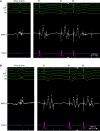
Examples of left ventricular EGM responses to RV extra-stimulation. (A) EDP site: during sinus rhythm (SR), a low-voltage EGM with fragmentation is recorded (left panel). During S1 (centre), two EGM components become visible: a low-frequency potential compatible with far-field and a high-frequency component compatible with near-field activity. During S2 (right), the near-field potential delays and splits into multiple components. (B) No EDP site: During SR, a low-voltage, fragmented EGM is recorded. During S1, two EGM components separate but do not delay further with the application of the extra-stimulus (S2). (C) EDP site: during SR, two EGM components can be distinguished: a high-voltage far-field EGM and a near-field EGM at the end of the QRS complex. During S1, no further delay of the near-field is appreciated, but it delays significantly during S2. (D) Bipolar EGMs recorded during SR and during RV pacing (S1 and S2) with a multipolar penta-spline multipolar catheter. During SR, fragmentation is observed in practically all bipoles. During S1, local near-field potentials become more evident in bipoles from 1–2 to 9–10, whereas bipoles from 13–14 to 19–20 show low-frequency potentials. During S2, near-field components delay and split further (EDPs) in consecutive bipoles from 1–2 to 9–10 but no delay is observed in bipoles from 13–14 to 19–20. The white triangles indicate the earliest and latest sharp peaks of the bipolar EGMs, and redt triangles indicate fragmented and delayed near-field potentials. EDP, evoked delayed potential; EGM, electrogram; RV, right ventricle.
Figure 1
Figure 1
Examples of left ventricular EGM responses to RV extra-stimulation. (A) EDP site: during sinus rhythm (SR), a low-voltage EGM with fragmentation is recorded (left panel). During S1 (centre), two EGM components become visible: a low-frequency potential compatible with far-field and a high-frequency component compatible with near-field activity. During S2 (right), the near-field potential delays and splits into multiple components. (B) No EDP site: During SR, a low-voltage, fragmented EGM is recorded. During S1, two EGM components separate but do not delay further with the application of the extra-stimulus (S2). (C) EDP site: during SR, two EGM components can be distinguished: a high-voltage far-field EGM and a near-field EGM at the end of the QRS complex. During S1, no further delay of the near-field is appreciated, but it delays significantly during S2. (D) Bipolar EGMs recorded during SR and during RV pacing (S1 and S2) with a multipolar penta-spline multipolar catheter. During SR, fragmentation is observed in practically all bipoles. During S1, local near-field potentials become more evident in bipoles from 1–2 to 9–10, whereas bipoles from 13–14 to 19–20 show low-frequency potentials. During S2, near-field components delay and split further (EDPs) in consecutive bipoles from 1–2 to 9–10 but no delay is observed in bipoles from 13–14 to 19–20. The white triangles indicate the earliest and latest sharp peaks of the bipolar EGMs, and redt triangles indicate fragmented and delayed near-field potentials. EDP, evoked delayed potential; EGM, electrogram; RV, right ventricle.
Figure 2
Figure 2
Procedural workflow. EDP, evoked delayed potential; LV, left ventricle; RV, right ventricle; VRP, ventricular refractory period; VT, ventricular tachycardia.
Figure 3
Figure 3
(A) Cumulative incidence of ventricular tachycardia recurrence after ablation. (B) Cumulative incidence of death, heart transplant, or LVAD implantation after ablation. HTx, heart transplantation; LVAD, left ventricular assist device; VT, ventricular tachycardia.
Figure 4
Figure 4
Cumulative incidence of ventricular tachycardia recurrence in centres with prior experience in EDP ablation vs. centres without prior experience in EDP ablation. EDP, evoked delayed potential; VT, ventricular tachycardia.
Figure 5
Figure 5
Acute and long-term ablation outcomes per centre. (A) Non-inducibility after ablation and (B) VT recurrence.

References

    1. Di Biase L, Burkhardt JD, Lakkireddy D, Carbucicchio C, Mohanty S, Mohanty P et al. Ablation of stable VTs versus substrate ablation in ischemic cardiomyopathy: the VISTA randomized multicenter trial. J Am Coll Cardiol 2015;66:2872–82. - PubMed
    1. Cronin EM, Bogun FM, Maury P, Peichl P, Chen M, Namboodiri N et al. 2019 HRS/EHRA/APHRS/LAHRS expert consensus statement on catheter ablation of ventricular arrhythmias. Europace 2019;21:1143–4. - PMC - PubMed
    1. Natale A, Zeppenfeld K, Della Bella P, Liu X, Sabbag A, Santangeli P et al. Twenty-five years of catheter ablation of ventricular tachycardia: a look back and a look forward. Europace 2023;25:euad225. - PMC - PubMed
    1. Silberbauer J, Oloriz T, Maccabelli G, Tsiachris D, Baratto F, Vergara P et al. Noninducibility and late potential abolition: a novel combined prognostic procedural end point for catheter ablation of postinfarction ventricular tachycardia. Circ Arrhythm Electrophysiol 2014;7:424–35. - PubMed
    1. Jaïs P, Maury P, Khairy P, Sacher F, Nault I, Komatsu Y et al. Elimination of local abnormal ventricular activities: a new end point for substrate modification in patients with scar-related ventricular tachycardia. Circulation 2012;125:2184–96. - PubMed

Publication types

MeSH terms