. 2025 Mar 11;19(9):8786-8798.
doi: 10.1021/acsnano.4c16192. Epub 2025 Feb 24.

Dynamic Assembly of Pentamer-Based Protein Nanotubes

Affiliations

Dynamic Assembly of Pentamer-Based Protein Nanotubes

Lukasz Koziej et al. ACS Nano. .

Abstract

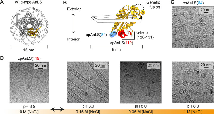
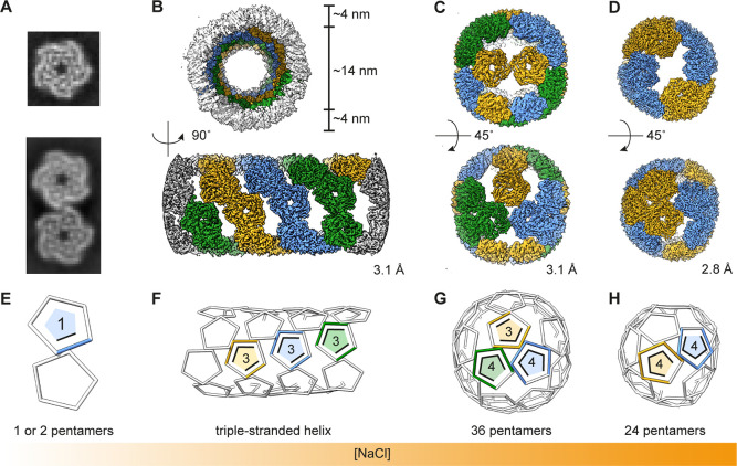
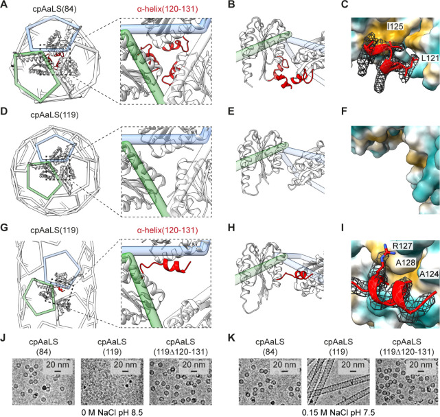
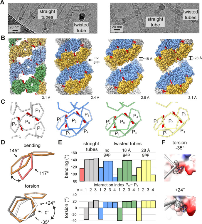
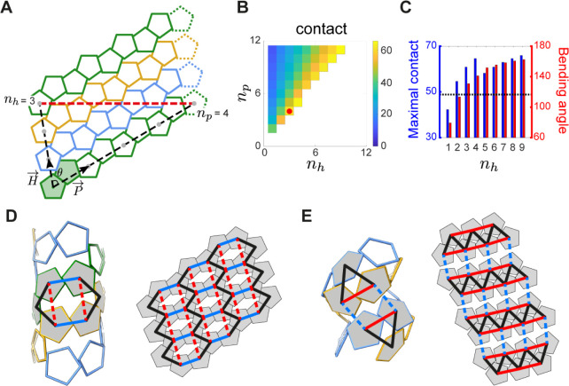
Hollow proteinaceous particles are useful nanometric containers for delivery and catalysis. Understanding the molecular mechanisms and the geometrical theory behind the polymorphic protein assemblies provides a basis for designing ones with the desired morphology. As such, we found that a circularly permuted variant of a cage-forming enzyme, Aquifex aeolicus lumazine synthase, cpAaLS, assembles into a variety of hollow spherical and cylindrical structures in response to changes in ionic strength. Cryogenic electron microscopy revealed that these structures are composed entirely of pentameric subunits, and the dramatic cage-to-tube transformation is attributed to the moderately hindered 3-fold symmetry interaction and the imparted torsion angle of the building blocks, where both mechanisms are mediated by an α-helix domain that is untethered from the native position by circular permutation. Mathematical modeling suggests that the unique double- and triple-stranded helical arrangements of subunits are optimal tiling patterns, while different geometries should be possible by modulating the interaction angles of the pentagons. These structural insights into dynamic, pentamer-based protein cages and nanotubes afford guidelines for designing nanoarchitectures with customized morphology and assembly characteristics.

Keywords: bionanotechnology; cryo-EM; geometry; non-quasi-equivalent; protein cage.


Conflict of interest statement

The authors declare no competing financial interest.

Figures

Figure 1
Figure 1
Assembly control of circularly permuted AaLS. (A) Structure of the dodecahedral wild-type AaLS cage (PDB ID 1HQK), shown as 12 wire pentagons with a ribbon diagram of a representative pentamer (gray) and protomer (orange). (B) Design of the circularly permuted variants, cpAaLS(84) and cpAaLS(119). The peptide linker connecting the native N- and C- termini (GTGGSGSS) is shown as a black dashed line. The new termini, C′(84) and N′(85) (blue) or C″(119) and N″(120) (red), are indicated by spheres. The α-helix(120–131), untethered by circular permutation for cpAaLS(119), is highlighted in red. (C,D) Cryo-EM micrographs of the cpAaLS(84) cage (C) and the NaCl- and pH-dependent cpAaLS(119) assemblies (D).
Figure 2
Figure 2
Cryo-EM structures of the cpAaLS(119) assemblies. (A–D) 2D classes (A) and 3D maps (B–D) of the cpAaLS assemblies, where colors (blue, orange, or green) indicate individual threads in the helical structure (B) or symmetry-related pentameric subunits in the spherical cages (C,D). The resolution of the final 3D reconstructions (GS-FSC at 0.143 cutoff) is provided at the right corner of each map. (E–H) The corresponding wire representation of the cpAaLS assemblies with the number of contacts per each asymmetric pentamer. Images are not to scale.
Figure 3
Figure 3
“Untethered” α-helix(120–131) facilitating the dynamic assembly of cpAaLS(119). (A) Wire diagram of the cpAaLS(84) assembly with an enlarged view of the 3-fold symmetry region. Three interacting monomers are shown as a ribbon with α-helix(120–131) highlighted in red. (B) Rotated side view of a pentamer pair (green and blue wire). The α-helix(120–131) domain from another monomer at the front is also shown to present the interaction at the 3-fold symmetry region in the cpAaLS(84) cage lumen. (C) Atomic interaction mode of the α-helix(120–131) domain in the cpAaLS(84) assembly. A unit is shown as a ribbon with amino acid side chains at the interface with the corresponding cryo-EM density map (mesh), and the interacting partners as hydrophilic (cyan) and hydrophobic (light brown) surfaces. (D–I) The corresponding representations for the cpAaLS(119) 24-pentameric spherical cage (D–F) and the tubular assembly (G–I), where the α-helix(120–131) is structurally disordered and was not modeled (D–F), or flipped to interact with an alternative surface (G–I), respectively. Panel (F) shows the same region as (C) to present the lack of cryoEM density corresponding to the α-helix(120–131) region. (J,K) Cryo-EM images of the cpAaLS(119) variant lacking α-helix(120–131), cpAaLS(119Δ120–131), compared to those of cpAaLS(84) and cpAaLS(119).
Figure 4
Figure 4
Capsomer interaction angles in the cpAaLS(119) tubes. (A) Cryo-EM micrographs of cpAaLS(119) (left) and cpAaLS(119, C37S, A85C) (right). (B) Cryo-EM maps of the straight tube composed of 3 evenly spaced helical strips (green, orange, and blue) and the twisted tubes featuring a variable gap (0, ∼18, or ∼28 Å) between dual strips (blue and orange). The resolution of the final 3D reconstructions (GS-FSC at 0.143 cutoff) is shown at the right corner of each map. The maps are not to scale. (C) The corresponding wire representations showing interactions of a pentamer (P0) with neighbors (P1–4). The α-helix(120–131) domains that accompany the intrathread interactions (P0–P2/3), otherwise invisible, are highlighted as red ribbons. (D,E) Conceptual representation (D) and the measured values (E) of the bending (top) and the torsion angles (bottom) between two pentamers. The wire diagrams show a pentamer–pentamer interaction in the cpAaLS(84) cage (red, top) and the cpAaLS(119) straight tubes (white and gray, bottom) with a reference having 0° torsion and 145° bending angles (brown). The interactions in the bar graph are colored and indexed as in (D), with those of the cpAaLS(84) cage (red bar) shown for comparison. (F) Two edges between interacting pentamers with the highest and the lowest torsion angles (−35° top, 24° bottom) observed in the cpAaLS(119) straight tube. The arginine side chain at the right vertex (R40, shown as gray sticks with mesh for the corresponding cryo-EM map) is flipped to maintain its interaction with a negatively charged patch (red surface) in the opposite pentamer.
Figure 5
Figure 5
Geometric rationale for the cpAaLS(119) tubular assembly. (A) A tiling representation of the cpAaLS(119) straight tube composed of three pentamer strips (green, blue, and orange). The lattice model is formed from periodic repeats of pentamer pairs (shaded in green) with vectors pointing along (P⃗) and across (H⃗) helical threads at an angle (θ). The tubular architecture is defined by the translation steps, helicity (nh) and periodicity (np) numbers, for two pentamers distanced by a single helical turn (gray dots connected by a black dashed line). The pentamers connected by the red dashed line are identical in the 3D tubular structure. (B) Heatmap presenting the percentage contact area between pentagonal edges in the tubular model for different (nh,np) combinations. The experimentally observed (nh,np) = (3,4) is indicated by a red dot. The structures in the blank area are geometrically or biologically forbidden. (C) The maximally possible contact area (blue) and the bending angle (red) for given nh over all the possible choices of np. The bending angle for the wild-type-like cpAaLS(84) cage is shown as a dotted line. (D,E) The interaction network rewiring between pentamers (shaded in gray) in the straight (D) and twisted tube (E), superimposed onto the 3D models (left) and the 2D tiling (right). Solid and dashed lines indicate contact and noncontact between pentamers, respectively. In the network transformation from the straight to the twisted tube, the blue contacts were lost, while the red ones were gained.

Similar articles

References

    1. Seeman N. C.; Sleiman H. F. DNA nanotechnology. Nat. Rev. Mater. 2017, 3 (1), 17068.10.1038/natrevmats.2017.68. - DOI
    1. Ulijn R. V.; Jerala R. Peptide and protein nanotechnology into the 2020s: Beyond biology. Chem. Soc. Rev. 2018, 47, 3391–3394. 10.1039/C8CS90055H. - DOI - PubMed
    1. Bale J. B.; Gonen S.; Liu Y.; et al. Accurate design of megadalton-scale two-component icosahedral protein complexes. Science 2016, 353, 389–394. 10.1126/science.aaf8818. - DOI - PMC - PubMed
    1. King N. P.; Bale J. B.; Sheffler W.; et al. Accurate design of co-assembling multi-component protein nanomaterials. Nature 2014, 510, 103–108. 10.1038/nature13404. - DOI - PMC - PubMed
    1. Golub E.; Subramanian R. H.; Esselborn J.; et al. Constructing protein polyhedra via orthogonal chemical interactions. Nature 2020, 578, 172–176. 10.1038/s41586-019-1928-2. - DOI - PMC - PubMed

Publication types

MeSH terms

Substances

LinkOut - more resources