. 2025 Feb;29(4):e70429.
doi: 10.1111/jcmm.70429.

Integrative Disulfidptosis-Based Risk Assessment for Prognostic Stratification and Immune Profiling in Glioma

Affiliations

Integrative Disulfidptosis-Based Risk Assessment for Prognostic Stratification and Immune Profiling in Glioma

Xiaowang Niu et al. J Cell Mol Med. 2025 Feb.

Abstract

Disulfidptosis, a new form of programmed cell death, plays a role in multiple types of cancer. However, research on disulfidptosis in glioma is lacking. A disulfidptosis-associated risk score was constructed using Cox regression modelling, while LASSO regression was applied for feature selection. To explore the relationship between the risk score and the immune microenvironment, we employed CIBERSORT, ssGSEA and ESTIMATE algorithms. Additionally, wet lab experiments were conducted to validate the functional role of the key disulfidptosis gene RPN1, demonstrating its ability to promote glioma cell proliferation and migration. Disulfidptosis genes were significantly upregulated in gliomas, influencing clinical features and survival. The risk score effectively predicted OS and varied among clinical subgroups. High-risk scores correlated with tumour growth, invasion and immunosuppression. Patients with different risk scores showed distinct immune cell infiltration patterns. Most immune checkpoints and chemokines were positively associated with risk scores. Laboratory findings confirmed that RPN1 significantly promoted glioma cell proliferation and migration. Disulfidptosis-based risk assessment stratifies glioma prognosis and reveals immune microenvironment characteristics, offering insights for personalised treatment strategies.

Keywords: disulfidptosis; glioma; immune microenvironment; prognosis; risk score.


Conflict of interest statement

The authors declare no conflicts of interest.

Figures
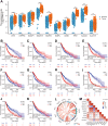
FIGURE 1
FIGURE 1
Expression and survival analysis of disulfide death gene in glioma. (A) mRNA expression levels of 10 disulfidptosis genes in the TCGA database were significantly upregulated in glioma. (B–K) Kaplan–Meier survival analysis of 10 disulfidptosis genes in glioma. (L–M) Correlation analysis of 10 disulfidptosis genes. TCGA, The Cancer Genome Atlas. *p < 0.05, ***p < 0.001.
FIGURE 2
FIGURE 2
Establishment of disulfidptosis genes‐associated risk scores. (A) Preliminary screening of 10 disulfidptosis genes by Univariate Cox regression analysis. (B) Selection plot of Lasso regression coefficients. (C) Plot of the trajectories of the Lasso regressors. (D) Multivariable Cox regression analysis to determine the established disulfidptosis genes‐associated risk score. (E) Time ROC analysis showed that the risk of glioma patients 1‐, 3‐ and 5‐year OS has good predictive value. (F) Risk factor map shows differences between high‐ and low‐risk score groups of patients' survival condition. RPN1, NDUFA11 and GYS1 show high expression in the high‐risk group, and NUBPL and LRPPRC show high expression in the low‐risk group (0: Alive, 1: Dead). (G) Kaplan–Meier survival analysis showed that patients with high‐risk scores have short lifetimes. (H) Kaplan–Meier survival analysis shows cumulative events of high‐risk score patients who have died. ROC, receiver operating characteristic.
FIGURE 3
FIGURE 3
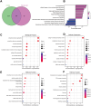
Enrichment analysis of differentially expressed genes between high–low‐risk groups. (A) The Venn diagram shows the intersection of differentially expressed genes in the TCGA and Rembrandt databases. (B) GSEA enrichment analysis. (C–E) GO function enrichment analysis. (F) KEGG pathway enrichment analysis. BP, biological process; CC, cellular components; GSEA, gene set enrichment analysis; KEGG, kyoto encyclopedia of genes and genomes; MF, molecular function; TCGA, The Cancer Genome Atlas.
FIGURE 4
FIGURE 4
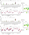
Relationship between risk score and tumour immune microenvironment. (A) In the TCGA database, the Cibersort algorithm showed significant differences in risk scores across 20 types of immunoinfiltrating cells. (B) The ssGSEA algorithm showed significant differences in risk scores among 19 types of immunoinfiltrating cells. (C) The estimate algorithm showed that the matrix scores, immune scores and estimated scores of the high‐risk group were higher than those of the low‐risk group. (D) In the CGGA database, the Cibersort algorithm showed significant differences in risk scores among 11 types of immunoinfiltrating cells. (E) The ssGSEA algorithm showed significant differences in risk scores across 20 types of immunoinfiltrating cells. (F) The Estimate algorithm also showed that patients in the high‐risk rating group had higher matrix scores, immune scores and estimated scores. CGGA, The Chinese Glioma Genome Atlas; TCGA, The Cancer Genome Atlas. *p < 0.05, **p < 0.01, ***p < 0.001.
FIGURE 5
FIGURE 5
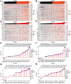
Risk scores can characterise immune checkpoint and chemokine expression levels in gliomas. (A and B) Coexpression heat maps of risk scores and 46 common immune checkpoints in the TCGA database. (C and D) Coexpression heat maps of risk scores and 42 common immune checkpoints in the CGGA database. (E and F) Lollipop charts of correlation between risk scores and 51 common chemokines in the TCGA database. (G and H) Lollipop chart of correlation between risk scores and 41 common chemokines in the CCGA database. CGGA, The Chinese Glioma Genome Atlas; ns, no signification; TCGA, The Cancer Genome Atlas. *p < 0.05, **p < 0.01, ***p < 0.001.
FIGURE 6
FIGURE 6
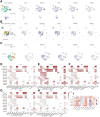
Relationship between five disulfidptosis genes and tumour immune infiltrating cells. (A–C) In TISCH database, the expression and distribution of five disulfidptosis genes in GSE89567, GSE131928 Smartseq2 and GSE70630. (D–H) In TISCH database, five disulfidptosis genes were expressed in 14 cell types in 13 single‐cell sequencing data sets. (I) Analysis of the correlation between five disulfidptosis genes and tumour purity and degree of immune cell infiltration in low‐grade gliomas and glioblastomas in TIMER database.
FIGURE 7
FIGURE 7
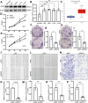
RPN1 significantly promoted the proliferation and migration of glioma cells. (A–C) The protein levels of RPN1 in U87, U251, U118 and U138 were significantly upregulated, which was consistent with the results in the CPTAC database. (D and E) The proliferation ability of U87 and U251 cells decreased after the RPN1 protein level was knocked down. (F–I) When RPN1 protein levels were knocked down, colony formation in U87 and U251 cells was reduced. (J–M) After knocking down RPN1 protein levels, the wound healing ability of U87 and U251 cells was reduced. (N–P) The migration ability of U87 and U251 cells was reduced after the RPN1 protein level was knocked down. *p < 0.05, **p < 0.01, ***p < 0.001, ****p < 0.0001.

References

    1. Zheng T., Liu Q., Xing F., Zeng C., and Wang W., “Disulfidptosis: A New Form of Programmed Cell Death,” Journal of Experimental & Clinical Cancer Research 42, no. 1 (2023): 137, 10.1186/s13046-023-02712-2. - DOI - PMC - PubMed
    1. Zheng P., Zhou C., Ding Y., and Duan S., “Disulfidptosis: A New Target for Metabolic Cancer Therapy,” Journal of Experimental & Clinical Cancer Research 42, no. 1 (2023): 103, 10.1186/s13046-023-02675-4. - DOI - PMC - PubMed
    1. Liu X., Nie L., Zhang Y., et al., “Actin Cytoskeleton Vulnerability to Disulfide Stress Mediates Disulfidptosis,” Nature Cell Biology 25, no. 3 (2023): 404–414, 10.1038/s41556-023-01091-2. - DOI - PMC - PubMed
    1. Machesky L. M., “Deadly Actin Collapse by Disulfidptosis,” Nature Cell Biology 25, no. 3 (2023): 375–376, 10.1038/s41556-023-01100-4. - DOI - PubMed
    1. Shao D., Shi L., and Ji H., “Disulfidptosis: Disulfide Stress Mediates a Novel Cell Death Pathway via Actin Cytoskeletal Vulnerability,” Molecules and Cells 46, no. 7 (2023): 414–416, 10.14348/molcells.2023.0060. - DOI - PMC - PubMed

MeSH terms

Substances