. 2026 Feb 1;21(2):612-635.
doi: 10.4103/NRR.NRR-D-24-01330. Epub 2025 Feb 24.

Neuronal guidance signaling in neurodegenerative diseases: Key regulators that function at neuron-glia and neuroimmune interfaces

Affiliations

Neuronal guidance signaling in neurodegenerative diseases: Key regulators that function at neuron-glia and neuroimmune interfaces

Junichi Yuasa-Kawada et al. Neural Regen Res. .

Abstract

The nervous system processes a vast amount of information, performing computations that underlie perception, cognition, and behavior. During development, neuronal guidance genes, which encode extracellular cues, their receptors, and downstream signal transducers, organize neural wiring to generate the complex architecture of the nervous system. It is now evident that many of these neuroguidance cues and their receptors are active during development and are also expressed in the adult nervous system. This suggests that neuronal guidance pathways are critical not only for neural wiring but also for ongoing function and maintenance of the mature nervous system. Supporting this view, these pathways continue to regulate synaptic connectivity, plasticity, and remodeling, and overall brain homeostasis throughout adulthood. Genetic and transcriptomic analyses have further revealed many neuronal guidance genes to be associated with a wide range of neurodegenerative and neuropsychiatric disorders. Although the precise mechanisms by which aberrant neuronal guidance signaling drives the pathogenesis of these diseases remain to be clarified, emerging evidence points to several common themes, including dysfunction in neurons, microglia, astrocytes, and endothelial cells, along with dysregulation of neuron-microglia-astrocyte, neuroimmune, and neurovascular interactions. In this review, we explore recent advances in understanding the molecular and cellular mechanisms by which aberrant neuronal guidance signaling contributes to disease pathogenesis through altered cell-cell interactions. For instance, recent studies have unveiled two distinct semaphorin-plexin signaling pathways that affect microglial activation and neuroinflammation. We discuss the challenges ahead, along with the therapeutic potentials of targeting neuronal guidance pathways for treating neurodegenerative diseases. Particular focus is placed on how neuronal guidance mechanisms control neuron-glia and neuroimmune interactions and modulate microglial function under physiological and pathological conditions. Specifically, we examine the crosstalk between neuronal guidance signaling and TREM2, a master regulator of microglial function, in the context of pathogenic protein aggregates. It is well-established that age is a major risk factor for neurodegeneration. Future research should address how aging and neuronal guidance signaling interact to influence an individual's susceptibility to various late-onset neurological diseases and how the progression of these diseases could be therapeutically blocked by targeting neuronal guidance pathways.

Keywords: TDP-43; TREM2; amyloid-β; axon guidance; neurodegeneration; neuroimmune interactions; neuroinflammation; neuron-glia interactions; neurovascular interactions; semaphorin; synaptic remodeling; tau; α-synuclein.


Conflict of interest statement

Conflicts of interest: The authors declare no conflicts of interest.

Figures

Figure 1
Figure 1
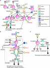
Roles of the neuronal guidance system under physiological and pathological conditions. The functionality of neuronal guidance signaling changes during developmental, adult, and neurodegenerative stages. (A) Axon midline crossing. Growing axons sense guidance cues and exhibit specific responses, such as attraction, repulsion, and cell adhesion. Their responsiveness can be switched in spatiotemporally regulated manners. For example, in the vertebrate neural tube, commissural axons initially extend toward the ventral midline by sensing netrin-1 (NTN1) derived from the floor plate or ventricular zone, which acts as an attractive or adhesion-promoting cue, respectively (Dominici et al., 2017; Moreno-Bravo et al., 2019; Wu et al., 2019). Upon reaching the midline, the axons lose their responsiveness to NTN1 and other attractants, including sonic hedgehog, and acquire responsiveness to repellents, such as SLIT, so that they smoothly cross the midline (Dickson and Zou, 2010; Yuasa-Kawada et al., 2023). (B) During prenatal and adult stages, synapses are remodeled to generate the mature neural architecture. Microglia-mediated synaptic pruning contributes to the refinement of the neural architecture. Locally externalized PtdSer (ePtdSer) is sensed as an “eat-me” signal by its receptors, such as TREM2 and GPR56, in microglia, and activates microglia to selectively engulf the “marked” presynaptic endings. Semaphorins (e.g., SEMA7A) may act as retrograde signals, to transduce neural activity and eliminate inappropriate presynaptic endings. (C) Aβ-exposed hyperactive presynaptic and postsynaptic structures are removed by activated microglia, via ePtdSer signaling. This mechanism exhibits beneficial effects during the early stages of neurodegeneration and possibly detrimental effects in the late stages (See Rueda-Carrasco et al., 2023 for details). (D and E) Cell contact-dependent SEMA-PLXN pathways mediate microglia-astrocyte and neuron-glia interactions and regulate glial activities under pathological conditions. (D) SEMA4D-PLXNB1 signaling induces the formation of peri-plaque glial nets by regulating glial cell spacing around Aβ plaques, promotes neuroinflammation, and affects microglial phagocytic behavior (see Huang et al., 2024 for details). Note that there is no verification to date whether the corresponding ligand is SEMA4D, although it is likely. (E) SEMA6D-PLXNA1-TREM2 signaling, which mediates interactions between Aβ-exposed excitatory neurons and microglia, induces microglial activation and promotes neuroinflammation around Aβ plaques (see Albanus et al., 2023 for details). Aβ: Amyloid-β; C: caudal; DAM: disease-associated microglia; FP: floor plate; PLXN: plexin; R: rostral; RP: roof plate; SEMA: semaphorin.
Figure 2
Figure 2
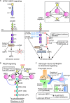
Networks of SEMA-PLXN and TREM2 signaling that mediate neuron-glia and neuroimmune interactions around disease-associated protein aggregates. Representative SEMAs, PLXNs, TREM2, and related signaling pathways in neurological diseases, including Alzheimer’s disease (AD) and amyotrophic lateral sclerosis (ALS), are depicted (see also Yuasa-Kawada et al., 2023, Figure 4). (A) Interactions of TREM2 and clusterin (CLU) with components of neuronal guidance signaling and abnormal protein aggregates, such as Aβ and TDP-43. TREM2 is involved in multiple diseases and may interact with multiple co-receptors. In (A), RND1 and RHOD-mediated control of the RAS/RAP-GAP activity of PLXNA4 is omitted. Risk factors for neurodegenerative diseases are shown in red. (B) SEMA6D-PLXNA1-TREM2 signaling. PLXNA1 and TREM2 form a receptor complex for SEMA6D and regulate microglial activation and function, including survival, proliferation, phagocytosis, and secretion of various cytokines. Additionally, the SEMA6D-PLXNA1 pathway utilizes both forward and reverse signaling. As shown in (A and B), TREM2 receives signals from a wide range of cues, such as Aβ, CLU, APOE, ePtdSer, and indirectly SEMA6D. (C) SEMA4D-PLXNB1/B2 signaling. PLXNB1 and PLXNB2 pathways seem to share most of their ligands and downstream signaling components. The RAS/RAP-GAP activity of PLXNB1 is regulated by RND1 and opposingly by RHOD (Liu et al., 2021). The SEMA6D-PLXNA1 pathway also utilizes both forward and reverse signaling. Such forward and reverse signaling pathways mediate bidirectional cell-cell communications in health and diseases, which can be general principles in neuronal guidance. APOE: Apolipoprotein E; APP: amyloid precursor protein; Aβ: amyloid-β; CLU: clusterin; DAP12: DNAX-activating protein of 12 kDa; GSK: glycogen synthase kinase; mTOR: mammalian target of rapamycin; NMDAR: N-methyl-D-aspartate-type glutamate receptor; NTN1: netrin; PI3K: phosphatidylinositol 3-kinase; PLXN: plexin; SEMA: semaphorin; TDP-43: transactive response DNA-binding protein 43 kDa; TLR: Toll-like receptor.
Figure 3
Figure 3
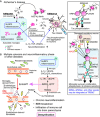
Neuroprotective and neuromodulatory roles of neuronal guidance signaling. (A) NTN1-UNC5 signaling affects Parkinson’s disease progression. NTN1-UNC5B/C signaling regulates the survival of dopamine neurons in the substantia nigra (SN), whereas NTN1 deficiency triggers α-synuclein (αSyn) aggregation and dopamine neuron death in Parkinson’s disease. The inset shows that NTN1 and RGMA share multiple receptors. A schematic illustration of guidance cues and receptors is based on domain structures annotated by EMBL-SMART (http://smart.embl-heidelberg.de/). (B) RELN signaling ameliorates AD progression. RELN upregulates GSK3B phosphorylation and suppresses GSK3B activity, which blocks tau phosphorylation. The rare variant RELN-H3447R acts as a gain-of-function (GOF) mutant to suppress tau phosphorylation more effectively than the major variants, even in AD families. Interestingly, RELN and APOE seem to competitively bind to VLDLR and APOER2. (C) Astrocytic EFNA3-neuronal EPHA4 forward and reverse signaling pathways control synaptic efficacy and remodeling. EFNA3-EPHA4 forward signaling triggers dendritic spine retraction, whereas EPHA4-EFNA3 reverse signaling reduces glutamate transporter levels in astrocytes, increasing intrasynaptic glutamate levels and synaptic efficacy. EFNA3 and NEO1 in astrocytes opposingly regulate the glutamate levels at synaptic spaces. AD: Alzheimer’s disease; AMPK: Adenosine 5′-monophosphate-activated protein kinase; APOE: apolipoprotein E; DD: death domain; GAP: GTPase-activating protein; GLAST: glial glutamate/aspartate transporter; GLT1: glutamate transporter subtype-1; Ig: immunoglobulin-like; LTP: long-term potentiation; NTN: netrin; PI3K: phosphatidylinositol 3-Kinase; PM: plasma membrane; RGMA: repulsive guidance molecule-a; TSP: thrombospondin type 1; UPA: UNC5-PIDD-ankirin; ZU5: present in ZO-1 and UNC5.
Figure 4
Figure 4
Evolutionarily conserved SARM-NMNAT-WNK machinery regulates axon degeneration, stabilization, and branching. (A) Signaling pathways of Wallerian degeneration in response to axon damage, driving axon destruction. Two enzymes, nicotinamide mononucleotide adenylyl-transferase 2 (NMNAT2), which synthesizes NAD+, and sterile α- and Toll/Interleukin-1 receptor motif-containing protein 1 (SARM1), which hydrolyzes NAD+, control Wallerian degeneration. The inset shows cycles of the metabolites and enzymes involved. NMN and NAD+ opposingly regulate SARM1 activity by binding to its armadillo repeat (ARM) domain. (B) Axonal fates, such as branching, stabilization, and degeneration, are controlled by the SARM-NMNAT-WNK mechanisms, in combination with AXED and MYCBP2, and possibly regulated by neuroguidance cues, including SEMA3A, RGMA, and SLIT. ADPR: ADP-ribose; NAD: nicotinamide adenine dinucleotide; NAM: nicotinamide; NMN: nicotinamide mononucleotide; NMNAT: nicotinamide mononucleotide adenylyltransferase; SEMA: semaphorin; WNK: with-no-lysine kinase.
Figure 5
Figure 5
Microglia-mediated neuroinflammatory responses and endothelial-to-mesenchymal transition (EndoMT) are regulated by neuronal guidance signaling. (A) Microglia-mediated neuroinflammation is triggered and maintained by SEMA6D-PLXNA1-TREM2 signaling in AD. Broad arrays of signals, including Aβ, SEMA6D, ePtdSer, CLU, and APOE, may be integrated at TREM2 to coordinate microglial responses, as shown on the right. SEMA3A-PLXNA4 signaling may promote Aβ-tau cross-seeding. It is uncertain whether PLXNA4 interacts with TREM2. (B) To prevent chronic neuroinflammation, BBB integrity is regulated by ARF6 signaling. BBB breakdown is driven by EndoMT, which is regulated by opposing actions of inflammatory cytokines (such as IL-1β and VEGFA) and neuroguidance cues (such as SLIT2). BBB breakdown leads to infiltration of immune cells into the brain parenchyma, and eventually to CNS demyelination, which is a clinical feature of multiple sclerosis. APOE: Apolipoprotein E; ARF: ADP ribosylation factor; Aβ: amyloid-β; BBB: blood–brain barrier; CLU: clusterin; DAP: DNAX-activating protein; GSK: glycogen synthase kinase; IL: interleukin; mTOR: mammalian target of rapamycin; NTN: netrin; PI3K: phosphoinositide 3-kinase; PLXN: plexin; PRR: pattern recognition receptor; SEMA: semaphorin; TNF: tumor necrosis factor; VEGFA: vascular endothelial growth factor A; VEGFR: vascular endothelial growth factor receptor.

References

    1. Adams RH, Eichmann A. Axon guidance molecules in vascular patterning. Cold Spring Harb Perspect Biol. 2010;2:a001875. - PMC - PubMed
    1. Ahn EH, Kang SS, Qi Q, Liu X, Ye K. Netrin1 deficiency activates MST1 via UNC5B receptor, promoting dopaminergic apoptosis in Parkinson’s disease. Proc Natl Acad Sci USA. 2020;117:24503–24513. - PMC - PubMed
    1. Akçimen F, Lopez ER, Landers JE, Nath A, Chiò A, Chia R, Traynor BJ. Amyotrophic lateral sclerosis: translating genetic discoveries into therapies. Nat Rev Genet. 2023;24:642–658. - PMC - PubMed
    1. Albanus RD, et al. Systematic analysis of cellular crosstalk reveals a role for SEMA6D-TREM2 regulating microglial function in Alzheimer’s disease. BioRxiv [preprint] 2023 doi: 10.1101/2022.11.11.516215.
    1. Alberti S, Hyman AA. Biomolecular condensates at the nexus of cellular stress, protein aggregation disease and ageing. Nat Rev Mol Cell Biol. 2021;22:196–213. - PubMed

LinkOut - more resources