. 2026 Apr 1;21(4):1383-1395.
doi: 10.4103/NRR.NRR-D-24-01016. Epub 2025 Feb 24.

Damage and repair in retinal degenerative diseases: Molecular basis through clinical translation

Affiliations

Damage and repair in retinal degenerative diseases: Molecular basis through clinical translation

Ziting Zhang et al. Neural Regen Res. .

Abstract

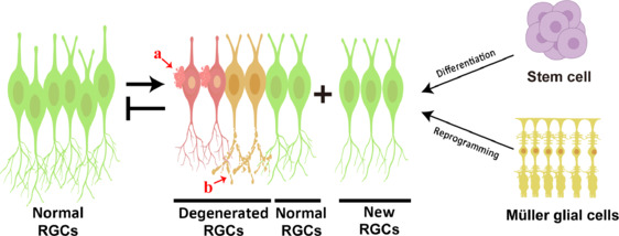
Retinal ganglion cells are the bridging neurons between the eye and the central nervous system, transmitting visual signals to the brain. The injury and loss of retinal ganglion cells are the primary pathological changes in several retinal degenerative diseases, including glaucoma, ischemic optic neuropathy, diabetic neuropathy, and optic neuritis. In mammals, injured retinal ganglion cells lack regenerative capacity and undergo apoptotic cell death within a few days of injury. Additionally, these cells exhibit limited regenerative ability, ultimately contributing to vision impairment and potentially leading to blindness. Currently, the only effective clinical treatment for glaucoma is to prevent vision loss by lowering intraocular pressure through medications or surgery; however, this approach cannot halt the effect of retinal ganglion cell loss on visual function. This review comprehensively investigates the mechanisms underlying retinal ganglion cell degeneration in retinal degenerative diseases and further explores the current status and potential of cell replacement therapy for regenerating retinal ganglion cells. As our understanding of the complex processes involved in retinal ganglion cell degeneration deepens, we can explore new treatment strategies, such as cell transplantation, which may offer more effective ways to mitigate the effect of retinal degenerative diseases on vision.

Keywords: cell replacement therapy; degeneration; glaucoma; optic nerve damage; regenerative medicine; retinal degenerative disease; retinal diseases; retinal ganglion cells; stem cell therapy; vision restoration.


Conflict of interest statement

Conflicts of interest: The authors declare no conflicts of interest.

Figures

Figure 1
Figure 1
Diagram of eyeballs status in health and common eye diseases. (A) In normal vision, light is focuses on the retina. (B) Glaucoma involves increased intraocular pressure damaging the optic nerve. (C) Diabetic Retinopathy results from retinal blood vessel damage due to high blood sugar, leading to hemorrhages and vision impairment. (D) Optic neuritis, often associated with autoimmune diseases, causes inflammation of the optic nerve, leading to vision loss and pain. (E) Traumatic optic neuropathy results in ocular or head trauma.
Figure 2
Figure 2
Mechanism diagram of RGCs damage and regeneration. Changes of various neurotrophic factors, a large number of reactive oxygen species, endoplasmic reticulum overstress, Ca2+ homeostasis imbalance, M1 microglia activation; The imbalance of mitochondrial division and fusion causes RGC degeneration. RGCs can be obtained from both male and female sources through several methods. ESCs can be isolated and cultured from human or mouse embryos of either gender. These ESCs can differentiate into RGCs when induced by a combination of Noggin, Dkk1, and IGF1. Similarly, iPSCs from male or female donors can be differentiated into RGCs using DLN (Dkk1 + Lefty A + Noggin) treatment and Atoh7 overexpression. Müller glial cells from both male and female donors can also be induced to differentiate into RGCs. BDNF: Brain-derived neurotrophic factor; CNTF: ciliary neurotrophic factor; EPO: erythropoietin; GDNF: glial cell-derived neurotrophic factor; IL-1β: interleukin-1β; NGF: nerve growth factor; PEDF: pigment epithelium-derived factor; RGCs: retinal ganglion cells; TNF-α: tumor necrosis factor-α; VEGF: vascular endothelial growth factor.
Figure 3
Figure 3
Timeline on RGC degeneration and regeneration. RGC degeneration and endogenous and exogenous factors promote RGC regeneration. CE: Ciliary epithelium; ESC: embryonic stem cell; EVs: extracellular vesicles; iPSCs: induced pluripotent stem cell; RGCs: retinal ganglion cells.
Figure 4
Figure 4
Changes in mitochondrial function under normal and pathological conditions. OPA1 and DRP1 are pivotal regulators of mitochondrial dynamics, executing opposing functions in the processes of fission and fusion. Research into the pathophysiology of glaucoma has illuminated the integral role these proteins play in maintaining mitochondrial functionality and vitality. It has been observed that the rate of mitochondrial division in the cellular and tissue structures of glaucoma patients markedly surpasses that of fusion. This increased incidence of mitochondrial division is not confined to human subjects alone; it is also evident in the RGCs and their axons in murine models of glaucoma pathology. DRP1: Dynamin-related protein 1; IOP: intraocular pressure; OPA1: optic atrophy type 1; RGCs: retinal ganglion cells.
Figure 5
Figure 5
RGCs damage and regeneration under pathological conditions. RGCs exhibit injury and axonal damage under pathological conditions. Stem cells and Müller glial cells can differentiate and reprogram into RGCs. “a” shows somatic damage, while “b” illustrates axonal injury. RGCs: Retinal ganglion cells.

References

    1. Agarwal D, Do H, Mazo KW, Chopra M, Wahlin KJ. Restoring vision and rebuilding the retina by Müller glial cell reprogramming. Stem Cell Res. 2023;66:103006. - PMC - PubMed
    1. Aguayo AJ, Rasminsky M, Bray GM, Carbonetto S, McKerracher L, Villegas-Pérez MP, Vidal-Sanz M, Carter DA. Degenerative and regenerative responses of injured neurons in the central nervous system of adult mammals. Philos Trans R Soc Lond B Biol Sci. 1991;331:337–343. - PubMed
    1. Ahmed A, Wang LL, Abdelmaksoud S, Aboelgheit A, Saeed S, Zhang CL. Minocycline modulates microglia polarization in ischemia-reperfusion model of retinal degeneration and induces neuroprotection. Sci Rep. 2017;7:14065. - PMC - PubMed
    1. Aires ID, Ribeiro-Rodrigues T, Boia R, Catarino S, Girão H, Ambrósio AF, Santiago AR. Exosomes derived from microglia exposed to elevated pressure amplify the neuroinflammatory response in retinal cells. Glia. 2020;68:2705–2724. - PubMed
    1. Akram R, Anwar H, Javed MS, Rasul A, Imran A, Malik SA, Raza C, Khan IU, Sajid F, Iman T, Sun T, Han HS, Hussain G. Axonal regeneration: underlying molecular mechanisms and potential therapeutic targets. Biomedicines. 2022;10:3186. - PMC - PubMed