Review
. 2025 Aug;67(8):986-1003.
doi: 10.1111/dmcn.16279. Epub 2025 Feb 25.

Cranial ultrasound in neonatal brain infections

Collaborators, Affiliations
Review

Cranial ultrasound in neonatal brain infections

Roosmarijn G Licht-van der Stap et al. Dev Med Child Neurol. 2025 Aug.

Abstract

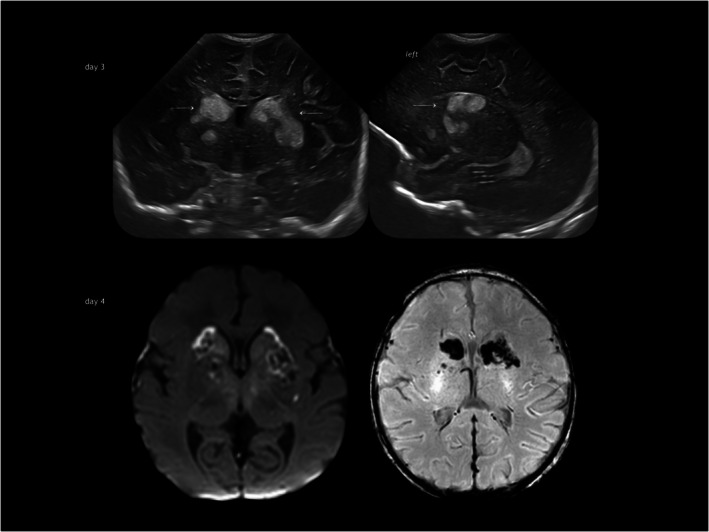
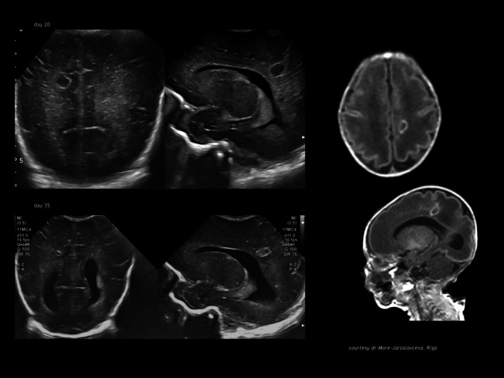
Infection of the neonatal central nervous system (CNS) can cause irreversible brain damage. Cranial ultrasound is an important neuroimaging modality in the neonatal period for detecting brain injury. Several types of organism can cause neonatal CNS infection. The aim of this narrative review is to provide an overview of the most common and typical ultrasonographic features of neonatal CNS infections and their evolution over time. Different microorganisms cause characteristic brain injury patterns. Using numerous imaging examples, we explain the different injury patterns caused by several Gram-positive and Gram-negative microorganisms, fungi, and viruses. This can guide the clinician to appropriate diagnosis and treatment.


Figures

FIGURE 1
FIGURE 1
The sequence of events in neonatal bacterial brain infection (coloured schemes are inverted images of actual patients).
FIGURE 2
FIGURE 2
Early‐onset group B Streptococcus meningitis: multiple arterial striatal perforator strokes in a term infant presenting with seizures and fever on postnatal day 6. Observe increase of echogenicity and sharper delineation of ischaemic areas in caudate head and putamen between day 6 and day 8 (arrows). T2‐weighted magnetic resonance image on day 9 illustrates the non‐haemorrhagic character of the juxtaventricular striatal lesions.
FIGURE 3
FIGURE 3
Early‐onset group B Streptococcus meningitis: ischaemic perforator strokes with haemorrhagic conversion (arrows) in an infant born at term with seizures and documented meningitis on day 3. Below the corresponding magnetic resonance image at day 4. Diffusion‐weighted imaging (left) and susceptibility‐weighted imaging (right) confirming the stroke lesions with haemorrhagic conversion.
FIGURE 4
FIGURE 4
Late‐onset group B Streptococcus meningitis: leptomeningitis with subcortical infarction. (a) Infant born late preterm with late‐onset GBS meningitis, prolonged antibiotic therapy; (sub)acute images: extensive subdural collections, arachnoiditis and meningeal thickening; cranial ultrasound at term (below) shows resolution of the subdural collections and ventricular dilatation which stabilized spontaneously afterwards. (b) Term‐born infant with late‐onset GBS meningitis: on day of admission hyperechoic inflammatory collections are suspected in and around the frontoparietal and occipital cortex, there is an increase of echogenicity in both thalami; the diffusion‐weighted MRI (right) confirms the stroke‐like lesions in the cortex and both thalami. Abbreviations: GBS, group B Streptococcus; MRI, magnetic resonance imaging; thal, thalami.
FIGURE 5
FIGURE 5
Escherichia coli meningitis and ventriculitis. (a) Infant born preterm (gestational age 26 weeks) with initial IVH grade 2, subsequent E. coli ventriculitis around day 5: evolving intraventricular changes, at first only faintly hyperechoic conglomerates, later adhesions (strands) and compartmentalization of the ventricle cavities. (b) Infant born preterm (gestational age 30 weeks) with initial IVH grade 3, subsequent E. coli ventriculitis around day 14 with abscess formation over the cerebellar surface as seen on a right mastoid fontanel view; observe dilatation of the lateral and third ventricles, sparing the fourth ventricle. (c) Infant born preterm (gestational age 35 weeks) with intraventricular strands, a collection of debris around the cerebellum on the mastoid fontanel view (arrow) and an enlarged foramen of Magendi and cisterna magna (pericerebellar arachnoid pathways hindered by inflammation). Abbreviations: CUS, cranial ultrasound; IVH, intraventricular haemorrhage; L, left; R, right.
FIGURE 6
FIGURE 6
Enterobacterial meningitis. (a) Infant born preterm (gestational age 32 weeks) with Enterobacter septicaemia, fasciotomy for necrosis of a foot, thrombocytopenia. On a midsagittal cranial ultrasound view from the anterior fontanel the upper vermis margin is obscured initially (arrow); mastoid insonation (middle) reveals intraparenchymal cerebellar destruction on serial imaging with membrane formation; the process ends in triventricular hydrocephalus necessitating placement of a reservoir for ventricular tapping (right image). (b) Infant born very preterm with Pseudomonas septicaemia and extensive microabscedation of the entire brain preceding total destruction before demise; this proceeds in less than 2 weeks. (c) Infant born preterm (gestational age 26 weeks), day 15: Serratia marcescens sepsis and meningitis; parenchymal circular hyperechoic change evolves into cavitation in 5 days. (d) Citrobacter encephalitis in a term infant: asymmetric, focal hyperechoic change of white matter and striatum evolves into extensive liquefactive necrosis of the entire parenchyma within a few days.
FIGURE 7
FIGURE 7
Bacillus cereus meningo‐encephalitis. (a) Infant born preterm (gestational age 30 weeks) with sepsis screen 48 hours before; lethargic, and full fontanelle but normal cerebrospinal fluid findings; cranial ultrasound shows predominantly unilateral extensive and irregular white matter echogenicity followed by destruction in the week after detection; the causative organism turned out to be Bacillus cereus. (b) Infant born preterm (33 weeks postmenstrual age) with Bacillus cereus in blood and cerebrospinal fluid: predominantly unilateral frontal white matter destruction.
FIGURE 8
FIGURE 8
Listeria encephalitis. (a) Medial cerebral artery stroke complicating Listeria septicaemia in a term infant with subsequent tissue loss on follow‐up imaging (4 weeks). (b) Bilateral parietal hyperechoic white matter change with radial character in listeria infection in an infant born preterm; observe additional inflammatory changes in plexus choroideus (arrow).
FIGURE 9
FIGURE 9
Staphylococcus aureus abscess. (a) Cerebral abscess in the paracentral lobule with S. aureus in a term infant (parasagittal cranial ultrasound image). (b) Cerebellar abscess with S. aureus (two different infants born preterm, both with mastoid insonations).
FIGURE 10
FIGURE 10
Aspergillus focal infection. HELLP syndrome, Caesarean section at gestational age 27 weeks, birthweight 600 g, skin infection at peripheral deep line on day 13, Aspergillus in blood and cerebrospinal fluid day 14; cranial ultrasound showing cyst in the right frontoparietal region. Magnetic resonance imaging result of 6‐week intravenous liposomal amphotericin B followed by voriconazole. Focal post‐inflammatory cavity in the right parietal subcortex.
FIGURE 11
FIGURE 11
Candida encephalitis with abscess formation. Infant born preterm (gestational age 29 weeks): acute stage of C. albicans encephalitis with disseminated hyperechoic nodules in the parenchyma and on choroid plexus; early macro‐abscess formation 1 week later due to confluence of several nodules.
FIGURE 12
FIGURE 12
Enterovirus encephalitis–meningitis. Infant born late preterm, seizures caused by enterovirus meningitis at 2 weeks; cranial ultrasound images showing early hyperechoic white matter change evolving into cavitation mainly in the frontal lobes. Observe bilaterality and near symmetry. Observe restricted diffusion in the corresponding periventricular areas and network injury in pulvinar on DW MRI in the acute stage (arrow) as well as restricted diffusion in the corpus callosum, optic radiation, and posterior limb of the internal capsule. Abbreviation: DW MRI, diffusion‐weighted magnetic resonance imaging.
FIGURE 13
FIGURE 13
Rotavirus encephalitis–meningitis. Infant born late preterm, seizures caused by rotavirus infection at 2 weeks after birth. Cranial ultrasound imaging showing initial hyperechoic change similar to flaring in preterm white matter injury, but ending in cavitation in the frontal areas (below). Right diffusion‐weighted magnetic resonance imaging showing the acute changes. Abbreviation: DW MRI, diffusion‐weighted magnetic resonance imaging.
FIGURE 14
FIGURE 14
Herpes simplex virus encephalitis. (a) Term‐born infant with the acute changes and total brain destruction on MRI, caused by HSV (courtesy L Ramenghi, Genova). (b) A term infant, seizures and fever day 28: thalamo‐cortical injury pattern of Herpes simplex virus infection; cranial ultrasound shows hyperechoic change in right thalamus, this is confirmed on ADC map MRI. Abbreviation: ADC, apparent diffusion coefficient; MRI, magnetic resonance imaging.

References

    1. Hallmaier‐Wacker LK, Andrews A, Hope R, Demirjian A, Lamagni TL, Collin SM. Incidence of infant Gram‐negative invasive bacterial infections in England, 2011‐2019: an observational study using population‐wide surveillance data. Arch Dis Child. 2023. Sep;108(9):762–767. - PubMed
    1. Okike IO, Johnson AP, Henderson KL, Blackburn RM, Muller‐Pebody B, Ladhani SN, et al. Incidence, etiology, and outcome of bacterial meningitis in infants aged. Clinical infectious diseases 2014;59(10):e150–e157. - PubMed
    1. Ouchenir L, Renaud C, Khan S, Bitnun A, Boisvert AA, McDonald J, et al. The epidemiology, management, and outcomes of bacterial meningitis in infants. Pediatrics. 2017. Jul;140(1):e20170476. - PubMed
    1. Stevens JP, Eames M, Kent A, Halket S, Holt D, Harvey D. Long term outcome of neonatal meningitis. Arch Dis Child Fetal Neonatal 2003;88:179–184. - PMC - PubMed
    1. Polin RA, Harris MC. Neonatal bacterial meningitis. Semin Neonatol 2001;6: 157–172. - PubMed

MeSH terms

LinkOut - more resources