. 2025 Feb 9;14(4):246.
doi: 10.3390/cells14040246.

BCL-B Promotes Lung Cancer Invasiveness by Direct Inhibition of BOK

Affiliations

BCL-B Promotes Lung Cancer Invasiveness by Direct Inhibition of BOK

Palaniappan Ramesh et al. Cells. .

Abstract

Expression of BCL-B, an anti-apoptotic BCL-2 family member, is correlated with worse survival in lung adenocarcinomas. Here, we show that BCL-B can mitigate cell death initiation through interaction with the effector protein BOK. We found that this interaction can promote sublethal mitochondrial outer membrane permeabilization (MOMP) and consequently generate apoptosis-flatliners, which represent a source of drug-tolerant persister cells (DTPs). The engagement of endothelial-mesenchymal-transition (EMT) further promotes cancer cell invasiveness in such DTPs. Our results reveal that BCL-B fosters cancer cell aggressiveness by counteracting complete MOMP.

Keywords: BCL-2 family; BCL-B; BOK; DTP; EMT; cancer; drug-resistance; invasiveness; mitochondrial permeabilization; persister phenotype.


Conflict of interest statement

The authors declare no conflicts of interest.

Figures

Figure 1
Figure 1
BCL-B is highly expressed in lung cancers and inhibits apoptosis in a BAX/BAK-independent manner. (A) BCL-B expression in lung adenocarcinoma (LUAD) and lung squamous cell cancer (LUSC) (T, red bars) compared to normal tissue (N, gray bars). * p < 0.05 (ANOVA). (B) Kaplan–Meier survival curve of patients with indicated cancers belonging to high or low BCL-B expression from the TCGA database (auto cut-off). p-values are indicated. (C) Fold change in the number of dead WT PC9 cells or (D) BAX BAK DKO PC9 cells treated with indicated drugs and with or without overexpression of BCL-B. Mean ± SD of n = 3 samples per condition are shown. Graphs represent one of three independent experiments. Two-way ANOVA with Šidák’s multiple comparisons test: *** p < 0.001; **** p < 0.0001; ns, not significant (p > 0.05).
Figure 2
Figure 2
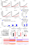
BCL-B inhibits BOK-dependent cell death. (A,B) Live cell imaging and detection of PI positive WT or BOK k.o. PC9 cells that have been treated with or without indicated drugs. Mean ± SD from n = 3 samples per group are shown. (C) Fold change in number of dead BAX BAK DKO PC9 cells treated with MG132 and with or without overexpression of BCL-B. Mean ± SD from n = 3 samples per condition are shown. Two-way ANOVA with Šidák’s multiple comparisons test: **** p < 0.0001; ns, not significant (p > 0.05). (D) Fold change in the number of dead BAX BAK BOK TKO PC9 cells treated with indicated drugs and with or without overexpression of BCL-B. Mean ± SD of n = 3 samples per condition are shown. Two-way ANOVA. (E) Fold change quantification of dead Bax Bak DKO MEF cells harboring doxycycline-inducible Bok, treated with indicated drugs with or without overexpression of murine Bcl-B (Diva). Mean ± SD of n = 3 samples per condition are shown. Two-way ANOVA with Šidák’s multiple comparisons test: *** p < 0.001; ns, not significant (p > 0.05). (F) Immunoblot with the indicated antibodies of cytosolic extracts from various indicated cell lines. (G) Fold change in the number of indicated cell lines silenced with either non-targeted control or BCL-B directed siRNAs before treatment with or without MG132. Mean ± SD of n = 3 samples per condition are shown. One-way ANOVA with Tukey’s multiple comparisons test: **** p < 0.0001; ** p < 0.01; * p < 0.05; ns, not significant (p > 0.05). (AG) All graphs represent one of three independent experiments. (H) Normalized BCL2 family gene expression in lung adenocarcinoma (LUAD) and lung squamous cell carcinoma (LUSC) of the TCGA pancancer cohort. Expression was normalized within LUAD and LUSC, respectively, following RNA-transcript processing using RSEM (RNA-Seq by Expectation-Maximization).
Figure 3
Figure 3
BCL-B binds to BOK. Co-immunoprecipitation experiments in cell lysates generated from HEK cells transfected with indicated flag-tagged anti-apoptotic human BCL-2 family members together with human BOK (hBOK) without (A) or with (B) treatment with MG132.
Figure 4
Figure 4
BCL-B promotes sublethal MOMP in DTPs. Fold change quantification of cytochrome c positive cells in control and DTP cells generated with MG132 (A) in WT PC9 or (B) BAX BAK DKO versus BAX BAK BOK TKO PC9 or (C) BOK k.o. PC9 cells or (D) indicated cell lines with overexpression of BCL-B. n = 6 samples are shown per condition. Unpaired t-test (A,C) or two-way ANOVA (B,D) with Šidák’s multiple comparisons test: **** p < 0.0001; ns, not significant (p > 0.05). All graphs represent one of three independent experiments.
Figure 5
Figure 5
EMT and cancer cell invasiveness is promoted in DTPs. (A) Live cell imaging analysis and (B) bottom transwell images of transmigrated BAX BAK DKO and BAX BAK BOK TKO PC9 control or DTP cells generated with MG132. Mean ± SD from n = 2 samples per group are shown. Counted cells are marked by blue mask. Scale bar 700µm. (C) Immunoblots of cell lysates from BAX BAK DKO and BAX BAK BOK TKO PC9 control or DTP cells treated with MG132 using indicated antibodies. (D) Incucyte analysis of transmigrated BAX BAK DKO and BAX BAK BOK TKO PC9 control or DTP cells generated with MG132, and treated with or without GM6001 20 µM. Mean ± SD from n = 2 samples per group are shown. All graphs represent one of three independent experiments.

References

    1. Bhola P., Letai A. Mitochondria—Judges and Executioners of Cell Death Sentences. Mol. Cell. 2016;61:695–704. doi: 10.1016/j.molcel.2016.02.019. - DOI - PMC - PubMed
    1. Garrido C., Galluzzi L., Brunet M., Puig P.E., Didelot C., Kroemer G. Mechanisms of Cytochrome c Release from Mitochondria. Cell Death Differ. 2006;13:1423–1433. doi: 10.1038/sj.cdd.4401950. - DOI - PubMed
    1. Tait S.W.G., Green D.R. Mitochondrial Regulation of Cell Death. CSH Perspect. Biol. 2013;5:a008706. doi: 10.1101/cshperspect.a008706. - DOI - PMC - PubMed
    1. Merino D., Kelly G.L., Lessene G., Wei A.H., Roberts A.W., Strasser A. BH3-Mimetic Drugs: Blazing the Trail for New Cancer Medicines. Cancer Cell. 2018;34:879–891. doi: 10.1016/j.ccell.2018.11.004. - DOI - PubMed
    1. Kalkavan H., Green D.R. MOMP, Cell Suicide as a BCL-2 Family Business. Cell Death Differ. 2018;25:46–55. doi: 10.1038/cdd.2017.179. - DOI - PMC - PubMed

Publication types

MeSH terms

Substances

LinkOut - more resources