. 2025 Feb 18;14(4):298.
doi: 10.3390/cells14040298.

Radiation Retinopathy: Microangiopathy-Inflammation-Neurodegeneration

Affiliations

Radiation Retinopathy: Microangiopathy-Inflammation-Neurodegeneration

Anja-Maria Davids et al. Cells. .

Abstract

Purpose: Proton irradiation is used to treat choroidal melanoma of the eye. The impact on non-malignant retinal cells is currently understudied. Therefore, we here report a mouse model to investigate the impact of proton irradiation on the retina.

Methods: We performed a proton beam irradiation of 5-15 Cobalt-Gray-Equivalent (CGE) of the eyes of female C57Bl6/J (Cx3cr1+/+), Cx3cr1gfp/+ and Cx3cr1gfp/gfp mice mimicking the clinical situation and evaluated the structure, function and cellular composition of the retina up to 24 weeks after irradiation.

Results: Proton beam irradiation of the eye with 15 CGE leads to cataract formation after 24 weeks without affecting the gross anatomy of the retinal vasculature as shown by Fundus imaging in all genotypes respectively. However, 10 and 15 CGE, lead to a significant decrease in NG2 positive cell numbers and all three dosages induced an increase in GFAP immunoreactivity. At 24 weeks a dosage of 15 CGE resulted in functional impairment and a decrease of NG2 positive cells in both WT and Cx3cr1 animals. Iba1 cell immunoreactivity was increased in all genotypes. However, in the Cx3cr1 animals the invasion of Iba1 cells into the deep vascular layer was partially prevented. This was accompanied by a less severe functional impairment in the irradiated Cx3cr1gfp/gfp vs. WT.

Conclusions: Although the gross anatomy of the retina does not seem to be affected by proton beam irradiation, the cellular composition and retinal function changed significantly in both WT and Cx3cr1 mice reflecting the clinical situation. Moreover, cataract formation was one of the major long-term effects of irradiation. We conclude that the murine model (WT and Cx3cr1 genotype) can be used to investigate proton-beam associated side effects in vivo as well as to test prospective interventions. Moreover, the loss of Cx3cr1 seems to be partially protective.

Keywords: radiation; retina; retinopathy.


Conflict of interest statement

The authors declare no conflict of interest.

Figures
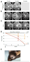
Figure 1
Figure 1
Vascular dynamics in the retina of the wild-type genotype after proton irradiation, time-course of fundus angiographies after proton irradiation in the irradiated eye (A), in the contralateral eye (B) and after sham procedure (C) as well as the development of cataract (D) with an exemplary photo (E).
Figure 2
Figure 2
Reduced number of pericytes in the irradiated retina (A) Immunofluorescence staining of pericytes in the central retinal flat mounts, Isolectin B4 (FITC) co NG2 (Cy3), of irradiated eyes (with 5, 10 and 15 CGE), upper row: deep vascular layer, * activated microglia, lower row superficial vascular layer, pericytes (white arrowhead). (B) Number of pericytes per mm vessel length in the retina, significantly decreased number of pericytes per mm in the irradiated eye in relation to the contralateral side and sham group for the doses 10 CGE (p < 0.001) and 15 CGE (p = 0.002).
Figure 3
Figure 3
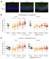
Gliosis in retinal sections of wild-type mice following irradiation (A) Immunofluorescence staining of central retinal sections of wild-type mice, GFAP (FITC) co Vimentin (Cy3), DAPI, of irradiated eyes (with 5, 10 and 15 CGE). (B) Relative integrated density of GFAP and Vimentin, irradiated versus contralateral side, GFAP: significant difference for every proton beam doses, no significance concerning sham (5 CGE: p < 0.001, 10 CGE: p = 0.007, 15 CGE: p < 0.001), Vimentin: no significant difference. (C) Relative integrated density of GFAP and Vimentin, central versus peripheral area, GFAP, Vimentin: no significant difference * p < 0.001.
Figure 4
Figure 4
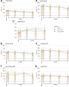
Comparison of Ganzfeld-ERG parameters regarding the genotype over time after proton irradiation with 15 CGE: (A) no significant difference between the genotypes, (B) no significant difference between the genotypes, (C) significant difference between the irradiated eyes of heterozygous Cx3cr1gfp/+ and wild-type mice (p = 0.003) or homozygous Cx3cr1gfp/gfp (p = 0.049), (D) tendency difference between the irradiated eyes of homozygous Cx3cr1gfp/gfp and heterozygous Cx3cr1gfp/+ mice (p = 0.072), (E) significant difference between the irradiated eyes of homozygous Cx3cr1gfp/gfp and heterozygous Cx3cr1gfp/+ mice) (p = 0.007) or wild-type mice (p = 0.043), (F) tendency difference between the irradiated eyes of homozygous Cx3cr1gfp/gfp and heterozygous Cx3cr1gfp/+ mice (p = 0.058), (G) significant difference between the irradiated eyes of homozygous Cx3cr1gfp/gfp and heterozygous Cx3cr1gfp/+ mice (p = 0.018).
Figure 5
Figure 5
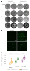
Activation of microglia following proton irradiation, (A) autofluorescence imaging of the retina of transgenic Cx3cr1gfp/gfp and Cx3cr1gfp/+ mice after proton irradiation, time-course of fundus autofluorescence’s before and 8, 16 as well as 24 weeks after proton irradiation in the irradiated and contralateral eye of Cx3cr1gfp/gfp and Cx3cr1gfp/+ mice, (B) immunofluorescence staining of Iba1 in retinal flat mounts of transgenic Cx3cr1gfp/+ and Cx3cr1gfp/gfp mice after proton irradiation with 15 CGE, Scale bar: 86 µm, (C) number of Iba1-positive cells per cubic millimeter, comparison of the genotypes significantly increased number of Iba1-positive cells in the retina of irradiated eyes of the wildtype (* p < 0.001), Cx3cr1gfp/gfp (* p < 0.001) and Cx3cr1gfp/+ mice (* p < 0.001) in relation to the contralateral side for the doses 15 CGE, no significant difference between the genotypes (p > 0.05).
Figure 6
Figure 6
Immunofluorescence staining of NG 2 and Isolectin B4 in retinal flat mounts of transgenic Cx3cr1gfp/+ (a–d) and Cx3cr1gfp/gfp mice after proton irradiation with 15 CGE, (A) upper row: deep vascular layer, lower row: superficial vascular layer (B) number of pericytes per mm vessel length 24 weeks after proton irradiation, comparison of the genotypes significantly decreased number of pericytes in the irradiated eye in relation to the contralateral side for the doses 15 CGE in Cx3cr1gfp/gfp (* p = 0.015), Cx3cr1gfp/+ (* p = 0.043) and wild-type mice (* p = 0.002); no difference between genotypes, tendency: wild-type mice have more pericytes per mm in contralateral eye than transgenic Cx3cr1 mice.
Figure 7
Figure 7
Gliosis in retinal sections of transgenic Cx3cr1gfp/+ and Cx3cr1gfp/gfp mice following irradiation, (A) Immunofluorescence staining of GFAP in central retinal sections of transgenic Cx3cr1gfp/+ (left) and Cx3cr1gfp/gfp mice (right) after proton irradiation with 15 CGE, contralateral non-irradiated versus irradiated retina, (B) relative integrated density of GFAP and Vimentin, all genotypes, irradiated versus contralateral side, GFAP: Increased integrated density with significant difference between the irradiated eye and the contralateral side for the Cx3cr1gfp/+ (* p = 0.016) and Cx3cr1gfp/gfp genotype (* p < 0.001), no significant difference between the genotypes (p > 0.05), Vimentin: no significant difference (p > 0.05), (C) relative integrated density of GFAP and Vimentin, central versus peripheral area GFAP, Vimentin: no significant difference.

References

    1. Ramos M.S., Echegaray J.J., Kuhn-Asif S., Wilkinson A., Yuan A., Singh A.D., Browne A.W. Animal models of radiation retinopathy—From teletherapy to brachytherapy. Exp. Eye Res. 2019;181:240–251. doi: 10.1016/j.exer.2019.01.019. - DOI - PubMed
    1. Egger E., Zografos L., Schalenbourg A., Beati D., Bohringer T., Chamot L., Goitein G. Eye retention after proton beam radiotherapy for uveal melanoma. Int. J. Radiat. Oncol. Biol. Phys. 2003;55:867–880. doi: 10.1016/S0360-3016(02)04200-1. - DOI - PubMed
    1. Fuss M., Loredo L.N., Blacharski P.A., Grove R.I., Slater J.D. Proton radiation therapy for medium and large choroidal melanoma: Preservation of the eye and its functionality. Int. J. Radiat. Oncol. Biol. Phys. 2001;49:1053–1059. doi: 10.1016/S0360-3016(00)01430-9. - DOI - PubMed
    1. Desjardins L., Levy C., d’Hermies F., Frau E., Schlienger P., Habrand J.L., Mammar H., Schwartz L., Mazal A., Delacroix S., et al. Initial results of proton therapy in choroidal melanoma at the d’Orsey Center for Proton Therapy; the first 464 cases. Cancer Radiother. 1997;1:222–226. doi: 10.1016/S1278-3218(97)89768-5. - DOI - PubMed
    1. Gragoudas E.S., Li W., Lane A.M., Munzenrider J., Egan K.M. Risk factors for radiation maculopathy and papillopathy after intraocular irradiation. Ophthalmology. 1999;106:1571–1577; discussion 1577–1578. doi: 10.1016/S0161-6420(99)90455-4. - DOI - PubMed

Substances

LinkOut - more resources