. 2025 Jan 29;47(2):86.
doi: 10.3390/cimb47020086.

Respiratory Muscle Injury Following Acute Monocled Cobra (Naja kaouthia) Envenoming: Histopathological Study in Rat Diaphragm

Affiliations

Respiratory Muscle Injury Following Acute Monocled Cobra (Naja kaouthia) Envenoming: Histopathological Study in Rat Diaphragm

Wanida Chuaikhongthong et al. Curr Issues Mol Biol. .

Abstract

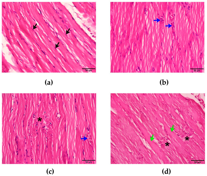
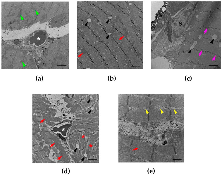
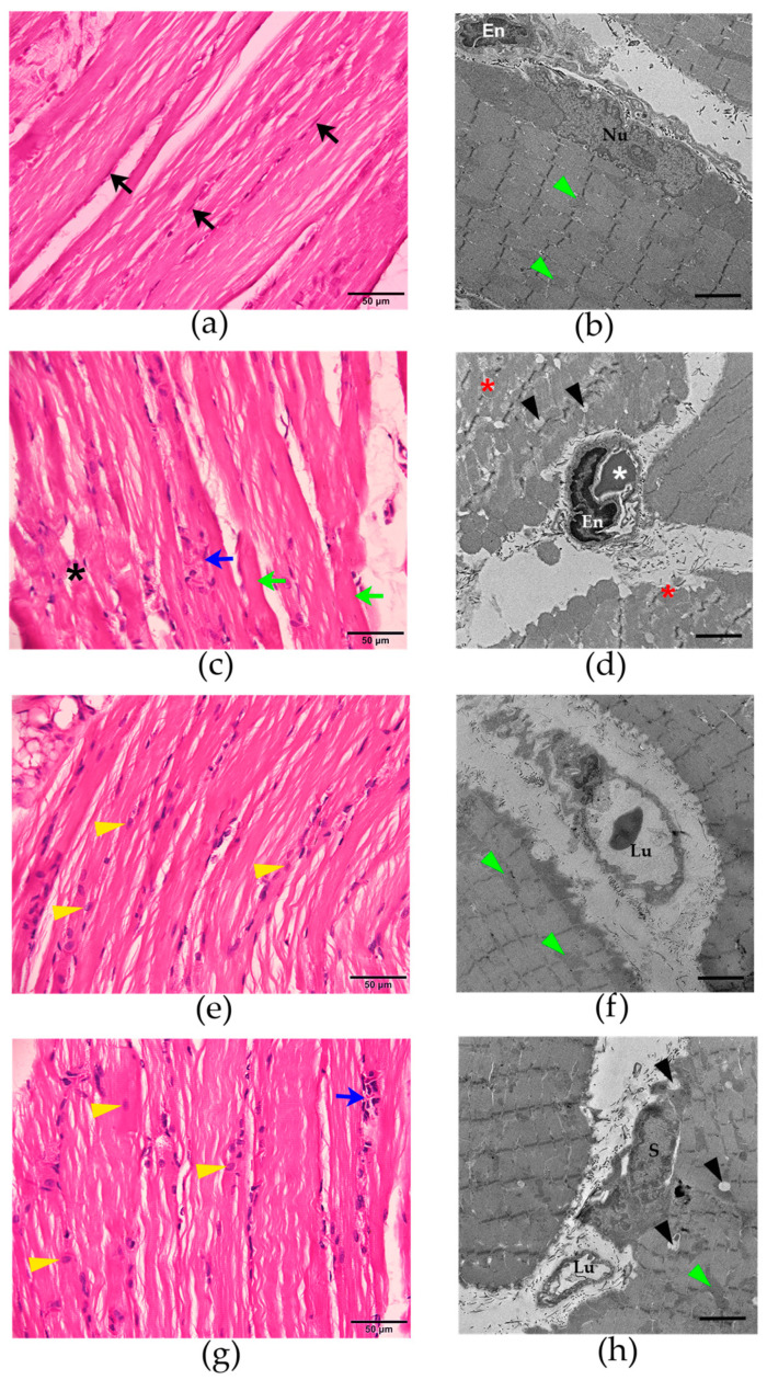
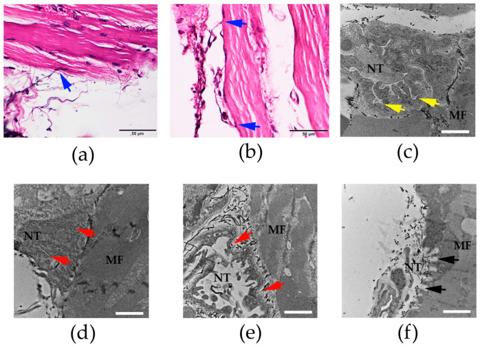
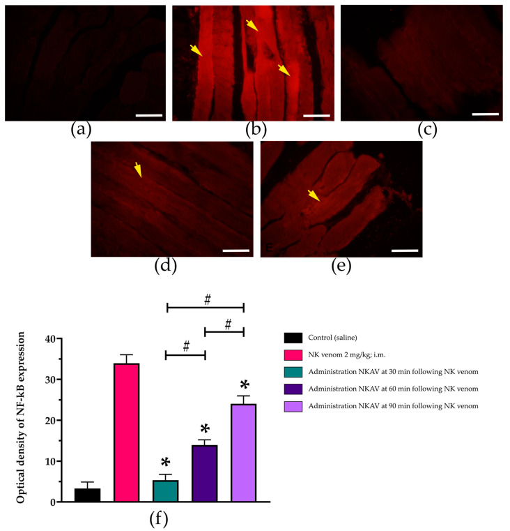
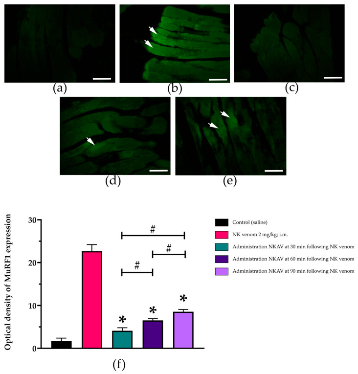
Clinical symptoms of monocled cobra (Naja kaouthia) envenoming include the paralysis of extraocular muscles, local tissue necrosis and death through respiratory failure. These neurotoxic outcomes are mainly due to the inhibitory action of postsynaptic neurotoxins to nicotinic acetylcholine receptors. However, injuries involving respiratory muscles have rarely been investigated. In this study, we determined the effect of N. kaouthia envenoming on morphological changes in the rat diaphragm. The efficacy of cobra monovalent antivenom in neutralising the histopathological effects of N. kaouthia venom was also evaluated. The intramuscular (i.m.) administration of N. kaouthia venom (2 mg/kg) caused skeletal muscle fibre atrophy and ruptures of myofibrils shown via a light microscope study. Transmission electron microscopy (TEM) revealed the zig-zagging of the Z-band, mitochondrial damages and degeneration of the synaptic fold of the neuromuscular junction following experimental cobra envenoming for 4 h. Intravenous administration of cobra antivenom at manufacturer-recommended doses diminished histopathological changes in the diaphragm following the administration of cobra venom. The expression of NF-kB and MuRF1 in the experimentally N. kaouthia-envenomed diaphragm indicated inflammation and tissue atrophy in the immunofluorescence analysis, respectively. In this study, we found that there were respiratory muscle injuries following N. kaouthia envenoming. The early administration of monovalent N. kaouthia antivenom is capable of neutralising neurotoxic outcomes following cobra envenoming.

Keywords: antivenom; diaphragm; histology; myotoxicity; snakebite.


Conflict of interest statement

The authors declare no conflicts of interest.

Figures

Figure 1
Figure 1
(a) Morphological changes of rat diaphragm after i.m. administration of saline (200 µL) showing the nucleus of the skeletal muscle cell (black arrows: H&E staining: scale bar = 50 µm). Morphological changes of rat diaphragm following administration of N. kaouthia venom (2 mg/kg) for (b) 60 and (c) 90 min showing red blood cell aggregations (blue arrow: H&E staining; scale bar = 50 µm) and myofibril ruptures (black asterisk: H&E staining; scale bar = 50 µm), respectively. (d) Morphological changes of rat diaphragm following the administration of N. kaouthia venom (2 mg/kg) for 4 h (H&E staining; scale bar = 50 µm: black asterisks indicate myofibril ruptures, green arrows indicate muscle hypertrophy).
Figure 2
Figure 2
(a) Transmission electron microscopy (TEM) image of diaphragm following the administration of saline shows the mitochondria (green arrowheads) and the red blood cells (white asterisk: scale bar = 2 µm). Morphological changes of rat diaphragm under TEM showing (b) mitochondrial swelling (red arrows: scale bar = 2 µm), intramyocellular lipid droplets (black arrowhead: scale bar = 2 µm) and (c) the zig-zagging of the Z-band (pink arrows: scale bar = 2 µm) following the administration of N. kaouthia venom (2 mg/kg, i.m.) for 90 min. (d) TEM image of rat diaphragm following administration of N. kaouthia venom (2 mg/kg) for 4 h showing a high degree of muscle fibre degeneration (red asterisks), mitochondrial swelling (red arrows), red blood cells (white asterisk) and intramyocellular lipid droplets (black arrowheads: scale bar = 2 µm). (e) TEM image of rat diaphragm showing mitochondrial degeneration (yellow arrowheads: scale bar = 2 µm) following the administration of N. kaouthia venom (2 mg/kg, i.m.) for 4 h. ‘S’ indicates the nucleus of satellite cells.
Figure 3
Figure 3
Morphological changes of the rat diaphragm after the i.m. administration of (a) saline (200 µL) showing the nucleus of the skeletal muscle cell (black arrows; H&E staining; scale bar = 50 µm) and (b) the mitochondria (green arrowheads; TEM image; scale bar = 2 µm). (c) Morphological changes of rat diaphragm (H&E staining; scale bar = 50 µm) following the administration of N. kaouthia venom (2 mg/kg: i.m.) for 4 h showing myofibril ruptures (black asterisks), red blood cell aggregations (blue arrow) and skeletal muscle hypertrophy (green arrows). (d) TEM image of rat diaphragm (scale bar = 2 µm) following the administration of N. kaouthia venom (2 mg/kg: i.m.) for 4 h shows high degree of muscle fibre degeneration (red asterisks), endothelial cell swelling (En), the appearances of red blood cells (white asterisk) and intramyocellular lipid droplets (black arrowheads). (e) H&E staining image and (f) TEM analysis (scale bar = 2 µm) of the rat diaphragm after i.v. administration of N. kaouthia monovalent antivenom (NKAV) for 30 min following experimental N. kaouthia envenomation (2 mg/kg, i.m.) show satellite cells (yellow arrowheads) and mitochondria (green arrowheads). (g) H&E staining image and (h) TEM analysis of the rat diaphragm after i.v. administration of NKAV at 90 min following the administration of N. kaouthia (2 mg/kg, i.m.) show the satellite cells (yellow arrowheads), red blood cell aggregations (blue arrow), intramyocellular lipid droplets (black arrowheads) and the mitochondria (green arrowheads). ‘Lu’ indicates the lumen of blood vessels. ‘En’ indicates endothelial cell swelling. ‘Nu’ indicates the nucleus. ‘S’ indicates the nucleus of satellite cells.
Figure 4
Figure 4
Morphological changes of the rat diaphragm (H&E staining; scale bar = 50 µm) after the i.m. administration of (a) saline (200 µL) and (b) N. kaouthia venom (2 mg/kg) showing the nerve terminal of nerve fibres (blue arrows). (c) Transmission electron microscopy image (TEM: scale bar = 2 µm) of the rat diaphragm following the administration of saline showing the synaptic folds of the neuromuscular junction (yellow arrows). (d) The administration of N. kaouthia venom (2 mg/kg: i.m.) for 90 min caused partial degeneration of the synaptic fold under TEM analysis (red arrows: scale bar = 2 µm). (e) TEM image of rat diaphragm shows severe degeneration and loss of the synaptic fold (red arrows) following i.m. administration of N. kaouthia venom (2 mg/kg) for 4 h. (f) The appearance of the synaptic fold (black arrows) in the rat diaphragm (TEM analysis: scale bar = 2 µm) after i.v. administration of NKAV at 30 min following the administration of N. kaouthia (2 mg/kg, i.m.). ‘NT’ indicates nerve terminal. ‘MF’ indicates muscle fibre.
Figure 5
Figure 5
Immunofluorescence analysis (scale bar = 50 µm) of the stimulation of NF-kB in the rat diaphragm tissues following the administration of (a) saline, (b) N. kaouthia venom (NK venom 2 mg/kg, i.m.), (c) N. kaouthia antivenom (NKAV, i.v.) following N. kaouthia venom (2 mg/kg, i.m.) at 30 min, (d) N. kaouthia antivenom (NKAV, i.v.) following N. kaouthia venom (2 mg/kg, i.m.) at 60 min and (e) N. kaouthia antivenom (NKAV, i.v.) following N. kaouthia venom (2 mg/kg, i.m.) at 90 min. (f) A comparison of optical density of NF-kB expression. Data represent the mean ± SEM (* p < 0.0001 compared with the group of NK venom (2 mg/kg, i.m.): # p < 0.0001, there was a significant difference when comparing between the groups, one-way analysis of variance (ANOVA); n = 3–5). Yellow arrows indicate the increase in NF-kB expression.
Figure 6
Figure 6
Immunofluorescence analysis (scale bar = 50 µm) of the stimulation of MuRF1 in the rat diaphragm tissues following the administration of (a) saline, (b) N. kaouthia venom (NK venom 2 mg/kg, i.m.), (c) NKAV (i.v.) following N. kaouthia venom (2 mg/kg, i.m.) at 30 min, (d) NKAV (i.v.) following N. kaouthia venom (2 mg/kg, i.m.) at 60 min and (e) NKAV (i.v.) following N. kaouthia venom (2 mg/kg, i.m.) at 90 min. (f) The comparison of optical density of MuRF1 expression. Data represent the mean ± SEM (* p < 0.0001 compared with NK venom (2 mg/kg, i.m.), # p < 0.0001, there was a significant difference when comparing between the groups, one-way ANOVA; n = 3–5). White arrows indicate the increase in MuRF1 expression.

Similar articles

References

    1. WHO . Guidelines for the Management of Snakebites. 2nd ed. WHO; Geneva, Switzerland: 2016. [(accessed on 10 September 2023)]. Available online: https://www.who.int/publications/i/item/9789290225300.
    1. Chanhome L., Cox M.J., Wilde H., Jintakoon P., Chaiyabutr N., Sitprija V. Venomous snakebite in Thailand I: Medically important snakes. Mil. Med. 1998;163:310–317. doi: 10.1093/milmed/163.5.310. - DOI - PubMed
    1. Shi S.C., Vogel G., Ding L., Rao D.Q., Liu S., Zhang L., Wu Z.-Y., Chen Z.N. Description of a New Cobra (Naja Laurenti, 1768; Squamata, Elapidae) from China with Designation of a Neotype for Naja atra. Animals. 2022;12:3481. doi: 10.3390/ani12243481. - DOI - PMC - PubMed
    1. WHO . Guidelines for the Management of Snake-Bites. WHO; Geneva, Switzerland: 2010. [(accessed on 5 December 2022)]. Available online: https://iris.who.int/bitstream/handle/10665/204464/B4508.pdf.
    1. Giri S., Taye S.J., Shyam R., Saikia B., Jangid R., Yasmin R., Doley R. Recurrent neurotoxity in Naja kaouthia envenomation: A case report from Assam, India. Toxicon. 2023;222:106990. doi: 10.1016/j.toxicon.2022.106990. - DOI - PubMed

LinkOut - more resources