. 2025 Jan 28;10(2):80.
doi: 10.3390/biomimetics10020080.

Biomechanical Optimization of the Human Bite Using Numerical Analysis Based on the Finite Element Method

Affiliations

Biomechanical Optimization of the Human Bite Using Numerical Analysis Based on the Finite Element Method

Maribel González-Martín et al. Biomimetics (Basel). .

Abstract

Biomechanical bite analysis is essential for understanding occlusal forces and their distribution, especially in the design and validation of dental prostheses. Although the finite element method (FEM) has been widely used to evaluate these forces, the existing models often lack accuracy due to simplified geometries and limited material properties.

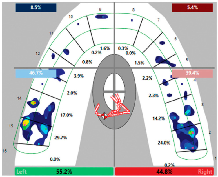
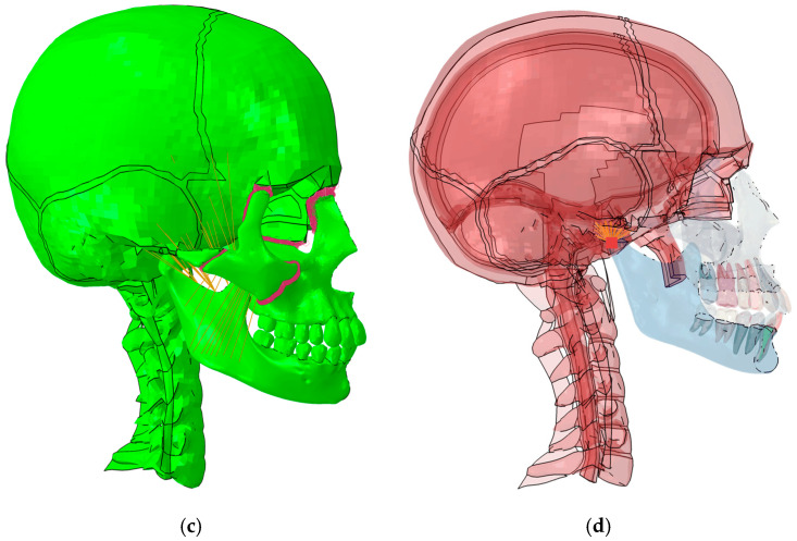
Methods: A detailed finite element model was developed using Abaqus Standard 2023 software (Dassault Systemes, Vélizy-Villacoublay, France), incorporating scanned 3D geometries of mandibular and maxillary bones. The model included cortical and cancellous bones (Young's modulus: 5.5 GPa and 13.7 GPa, respectively) and was adjusted to simulate bite forces of 220.7 N based on experimental data. Occlusal forces were evaluated using flexible connectors that replicate molar-to-molar interactions, and the stress state was analyzed in the maxillary and mandibular bones.

Results: The FEM model consisted of 1.68 million elements, with mesh sizes of 1-1.5 mm in critical areas. Bite forces on the molars were consistent with clinical trials: first molar (59.3 N), second molar (34.4 N), and third molar (16.7 N). The results showed that the maximum principal stresses in the maxillary bones did not exceed ±5 MPa, validating the robustness of the model for biomechanical predictions.

Conclusion: The developed model provides an accurate and validated framework for analyzing the distribution of occlusal forces in intact dentures. This approach allows the evaluation of complex prosthetic configurations and their biomechanical impact, optimizing future designs to reduce clinical complications and improve long-term outcomes. The integration of high-resolution FEM models with clinical data establishes a solid foundation for the development of predictive tools in restorative dentistry.

Keywords: bite force; dental biomechanics; dental prosthesis; finite element method; numerical simulation; occlusal force distribution.


Conflict of interest statement

The authors declare no conflicts of interest.

Figures

Figure 1
Figure 1
Distribution of bite pressures from the article by Turkistani et al [1]. The colour scale (not included in the reference) is defined from blue to red, where red indicates the highest pressure.
Figure 2
Figure 2
FEM model of bite analysis with intact dentition. (a) General lateral view; (b) detail of lateral view; (c) Tie-type kinematic constraints; (d) temporomandibular joint. Different colours have been defined for clarity, and only represent different parts of the model skull. The only exception is image c, where the purple colour represents the articulation between the bones using Tie-type kinematic constraints. In addition, image d has been updated to better show the point of the temporomandibular joint.
Figure 2
Figure 2
FEM model of bite analysis with intact dentition. (a) General lateral view; (b) detail of lateral view; (c) Tie-type kinematic constraints; (d) temporomandibular joint. Different colours have been defined for clarity, and only represent different parts of the model skull. The only exception is image c, where the purple colour represents the articulation between the bones using Tie-type kinematic constraints. In addition, image d has been updated to better show the point of the temporomandibular joint.
Figure 3
Figure 3
Model of the interaction between teeth. Green is for solid FEM elements. Red for the spring element that model the interaction and yellow for the distributing coupling that distributes the interaction force to the tooth surface.
Figure 4
Figure 4
Forces exerted by maxillary muscles and tendons.
Figure 5
Figure 5
Occlusion forces in 220.7 N bite.
Figure 6
Figure 6
Stress state: (a) maximum principal stresses [N/mm2]; (b) minimum principal stresses [N/mm2].

References

    1. Turkistani K.A., Alkayyal M.A., Abbassy M.A., Al-Dharrab A.A., Zahran M.H., Melis M., Zawawi K.H. Comparison of occlusal bite force distribution in subjects with different occlusal characteristics. Cranio. 2023;41:204–211. doi: 10.1080/08869634.2020.1830662. - DOI - PubMed
    1. Vaidyanathan A.K., Banu R.F. Finite element analysis—Concepts for knowledge and implementation in dental research. J. Indian Prosthodont. Soc. 2022;22:211–214. doi: 10.4103/jips.jips_299_22. - DOI - PMC - PubMed
    1. Zhou T., Wongpairojpanich J., Sareethammanuwat M., Lilakhunakon C., Buranawat B. Digital occlusal analysis of pre and post-single posterior implant restoration delivery: A pilot study. PLoS ONE. 2021;16:e0252191. doi: 10.1371/journal.pone.0252191. - DOI - PMC - PubMed
    1. Kim Y., Oh T.J., Misch C.E., Wang H.L. Occlusal considerations in implant therapy: Clinical guidelines with biomechanical rationale. Clin. Oral. Implants Res. 2005;16:26–35. doi: 10.1111/j.1600-0501.2004.01067.x. - DOI - PubMed
    1. Carl M. Dental Implant Prosthetics. Elsevier; Amsterdam, The Netherlands: 2015. Occlusal Considerations for Implant-Supported Prostheses; pp. 874–912.

LinkOut - more resources