. 2025 Jan 29;11(2):102.
doi: 10.3390/jof11020102.

Proteomic Approach to Study the Effect of Pneumocystis jirovecii Colonization in Idiopathic Pulmonary Fibrosis

Affiliations

Proteomic Approach to Study the Effect of Pneumocystis jirovecii Colonization in Idiopathic Pulmonary Fibrosis

Jonás Carmona-Pírez et al. J Fungi (Basel). .

Abstract

Idiopathic pulmonary fibrosis (IPF) is a chronic, progressive, and interstitial disease with an unclear cause, believed to involve genetic, environmental, and molecular factors. Recent research suggested that Pneumocystis jirovecii (PJ) could contribute to disease exacerbations and severity. This article explores how PJ colonization might influence the pathogenesis of IPF. We performed a proteomic analysis to study the profile of control and IPF patients, with/without PJ. We recruited nine participants from the Virgen del Rocio University Hospital (Seville, Spain). iTRAQ and bioinformatics analyses were performed to identify differentially expressed proteins (DEPs), including a functional analysis of DEPs and of the protein-protein interaction networks built using the STRING database. We identified a total of 92 DEPs highlighting the protein vimentin when comparing groups. Functional differences were observed, with the glycolysis pathway highlighted in PJ-colonized IPF patients; as well as the pentose phosphate pathway and miR-133A in non-colonized IPF patients. We found 11 protein complexes, notably the JAK-STAT signaling complex in non-colonized IPF patients. To our knowledge, this is the first study that analyzed PJ colonization's effect on IPF patients. However, further research is needed, especially on the complex interactions with the AKT/GSK-3β/snail pathway that could explain some of our results.

Keywords: Pneumocystis colonization; Pneumocystis jirovecii; iTRAQ quantification; idiopathic pulmonary fibrosis; protein–protein interaction networks; proteomics.


Conflict of interest statement

The authors declare no conflicts of interest.

Figures
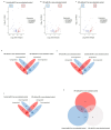
Figure 1
Figure 1
Differentially expressed proteins (DEPs) associated with the groups: control with Pneumocys jirovecii (PJ), idiopathic pulmonary fibrosis (IPF) colonized by PJ, and IPF non-colonized by PJ (AC). The DEPs were identified by comparing to the control group non-colonized by PJ; the comparison results are presented as volcano plots. The protein expression differences with a fold change of 0.8 or 1.2 (cutoff line, x = 0.260344) were considered significant at a p value of 0.05 (cutoff line, y = 1.30). The numbers in the colored frames indicate the identified DEP numbers. The dots that are labeled with protein designations represent some DEPs in the study. (DF) Overlapping DEPs were analyzed, and the shared protein numbers between the groups are shown as a Venn diagram. (G) Numbers of shared proteins among the groups are shown as a Venn diagram.
Figure 2
Figure 2
Pathway and process enrichment analysis associated with the groups: control with Pneumocystis jirovecii (PJ), idiopathic pulmonary fibrosis (IPF) colonized by PJ, and IPF non-colonized by PJ (AC). We performed a functional analysis for each given gene list, in the following ontology sources: GO Biological Processes, Reactome Gene Sets (REAC), KEGG pathways, WikiPathways (WP) and MIRNA. It includes up to 5 terms in each category and ordered by −log10 p-value. We built a protein-protein interaction network for each gene list (respectively, (D,E,F)) using the STRING database. We applied the Molecular Complex Detection (MCODE) algorithm to identify densely connected protein complexes. Then, we performed a functional analysis as described previously. The complexes found were named considering the best-scoring terms and the consensus of all researchers.

References

    1. Lederer D.J., Martinez F.J. Idiopathic Pulmonary Fibrosis. N. Engl. J. Med. 2018;378:1811–1823. doi: 10.1056/NEJMra1705751. - DOI - PubMed
    1. Althobiani M.A., Russell A.M., Jacob J., Ranjan Y., Folarin A.A., Hurst J.R., Porter J.C. Interstitial Lung Disease: A Review of Classification, Etiology, Epidemiology, Clinical Diagnosis, Pharmacological and Non-Pharmacological Treatment. Front. Med. 2024;11:1296890. doi: 10.3389/fmed.2024.1296890. - DOI - PMC - PubMed
    1. Ley B., Collard H.R., King T.E. Clinical Course and Prediction of Survival in Idiopathic Pulmonary Fibrosis. Am. J. Respir. Crit. Care Med. 2011;183:431–440. doi: 10.1164/rccm.201006-0894CI. - DOI - PubMed
    1. Muri J., Durcová B., Slivka R., Vrbenská A., Makovická M., Makovický P., Škarda J., Delongová P., Kamarád V., Vecanová J. Idiopathic Pulmonary Fibrosis: Review of Current Knowledge. Physiol. Res. 2024;73:487–497. doi: 10.33549/physiolres.935322. - DOI - PMC - PubMed
    1. Moore B.B., Moore T.A. Viruses in Idiopathic Pulmonary Fibrosis Etiology and Exacerbation. Ann. Am. Thorac. Soc. 2015;12:S186–S192. doi: 10.1513/AnnalsATS.201502-088AW. - DOI - PMC - PubMed

LinkOut - more resources