

Conformation and Membrane Topology of the N-Terminal Ectodomain of Influenza A M2 Protein
- PMID: 39997666
- PMCID: PMC11857740
- DOI: 10.3390/membranes15020040
Conformation and Membrane Topology of the N-Terminal Ectodomain of Influenza A M2 Protein
Abstract
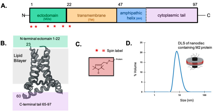
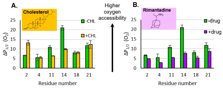
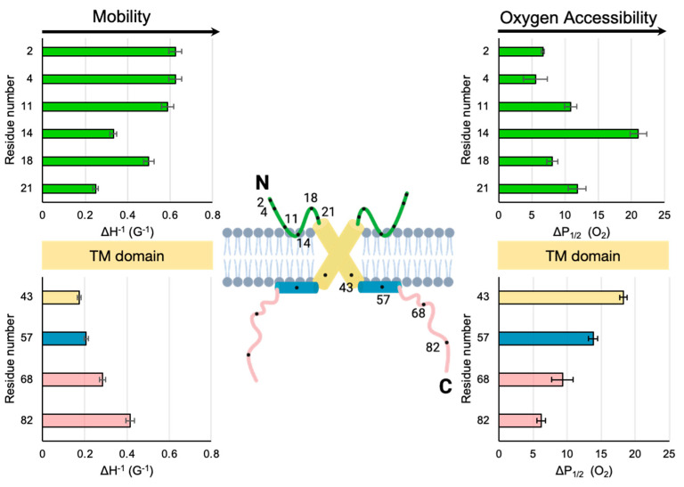
The N-terminal ectodomain of the influenza A M2 protein is a target for universal influenza vaccine development and novel antiviral strategies. Despite the significance of this domain, it is poorly understood and most structural studies of the M2 protein have disregarded the N-terminal ectodomain in their analyses. Here, we report conformational properties and describe insights into the membrane topology of sites along the N-terminal ectodomain. Full-length M2 protein is embedded in lipid bilayer nanodiscs and studied using site-directed spin labeling electron paramagnetic resonance spectroscopy. Results are consistent with a turn in the middle of the ectodomain that changes in proximity to the membrane surface upon the addition of cholesterol or the antiviral drug rimantadine. Similarly to other domains of M2 protein, lineshape analysis suggests that the N-terminal ectodomain can adopt multiple conformations.
Keywords: EPR; M2 protein; ectodomain; influenza; spin labels; universal vaccine.
Conflict of interest statement
The authors declare no conflicts of interest.
Figures

Similar articles
-
Conformation of influenza AM2 membrane protein in nanodiscs and liposomes.Biochim Biophys Acta Biomembr. 2023 Jun;1865(5):184152. doi: 10.1016/j.bbamem.2023.184152. Epub 2023 Mar 21. Biochim Biophys Acta Biomembr. 2023. PMID: 36948480 Free PMC article.
-
The Influenza M2 Ectodomain Regulates the Conformational Equilibria of the Transmembrane Proton Channel: Insights from Solid-State Nuclear Magnetic Resonance.Biochemistry. 2016 Sep 27;55(38):5387-97. doi: 10.1021/acs.biochem.6b00727. Epub 2016 Sep 12. Biochemistry. 2016. PMID: 27571210 Free PMC article.
-
The distal cytoplasmic tail of the influenza A M2 protein dynamically extends from the membrane.Biochim Biophys Acta Biomembr. 2019 Aug 1;1861(8):1421-1427. doi: 10.1016/j.bbamem.2019.05.021. Epub 2019 May 30. Biochim Biophys Acta Biomembr. 2019. PMID: 31153909 Free PMC article.
-
EPR Lineshape Analysis to Investigate the SNARE Folding Intermediates.Methods Mol Biol. 2019;1860:33-51. doi: 10.1007/978-1-4939-8760-3_3. Methods Mol Biol. 2019. PMID: 30317497 Free PMC article. Review.
-
Elucidating the design principles of photosynthetic electron-transfer proteins by site-directed spin labeling EPR spectroscopy.Biochim Biophys Acta. 2016 May;1857(5):548-556. doi: 10.1016/j.bbabio.2015.08.009. Epub 2015 Sep 1. Biochim Biophys Acta. 2016. PMID: 26334844 Review.
References
-
- Kandeil A., Patton C., Jones J.C., Jeevan T., Harrington W.N., Trifkovic S., Seiler J.P., Fabrizio T., Woodard K., Turner J.C., et al. Rapid evolution of A(H5N1) influenza viruses after intercontinental spread to North America. Nat. Commun. 2023;14:3082. doi: 10.1038/s41467-023-38415-7. - DOI - PMC - PubMed
Grants and funding
LinkOut - more resources
Full Text Sources
