. 2025 Feb 13;17(2):86.
doi: 10.3390/toxins17020086.

Thermal Stability and Matrix Binding of Citrinin in the Thermal Processing of Starch-Rich Foods

Affiliations

Thermal Stability and Matrix Binding of Citrinin in the Thermal Processing of Starch-Rich Foods

Lea Brückner et al. Toxins (Basel). .

Abstract

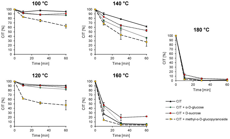
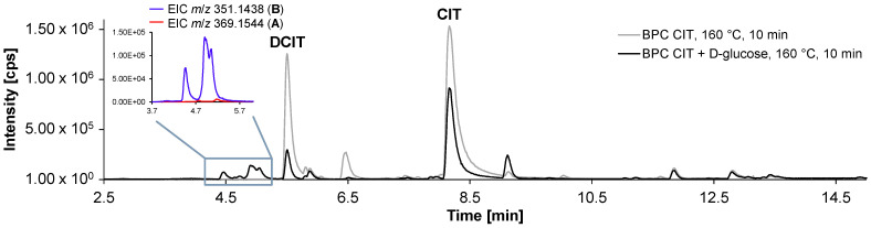
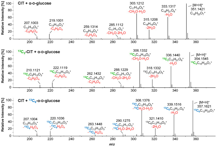
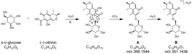
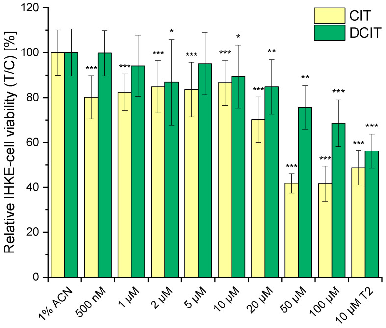
Citrinin (CIT) is a nephrotoxic mycotoxin commonly found in a broad range of foods, including cereals, spices, nuts, or Monascus fermentation products. Analyses have shown that CIT is present in processed foods in significantly lower concentrations than in unprocessed materials. Modified forms of CIT arising during food processing may provide an explanation for the discrepancy. This study deals with the thermal stability of CIT and the formation of reaction products of CIT with carbohydrates, followed by toxicological evaluations using cell culture models. HPLC-HRMS degradation curves of CIT heated in different matrix model systems were recorded, and the formation of decarboxycitrinin (DCIT), the main degradation product, was quantified. Additionally, chemical structures of reaction products of CIT with carbohydrates were tentatively identified using MS/MS spectra and stable isotope labelling. Subsequently, the degradation of CIT during biscuit baking was studied, and carbohydrate-bound forms of CIT were detected after enzymatic starch digestion. The formation of DCIT could explain the majority of CIT degradation, but, depending on the process, covalent binding to carbohydrates can also be highly relevant. Cytotoxicity of DCIT in IHKE-cells was found to be lower compared to CIT, while the toxicity as well as the intestinal metabolism of carbohydrate-bound CIT was not evaluated.

Keywords: citrinin; degradation; food; matrix binding; modified mycotoxin; mycotoxin.


Conflict of interest statement

The authors declare no conflicts of interest.

Figures

Figure 1
Figure 1
Chemical structure of CIT in its tautomeric forms p-quinone and o-quinone (3:2 equilibrium between the p- and o-tautomers in the solid state at room temperature), as well as the chemical structure of the 13C3-labeled CIT.
Figure 2
Figure 2
Degradation curves representing the thermal stability of CIT during heating for different times (10, 30, 60 min) and temperatures (100, 120, 140, 160, 180 °C) with and without the addition of model compounds to mimic different carbohydrates (α-d-glucose; d-sucrose; methyl-α-d-glucopyranoside). Methyl-α-d-glucopyranoside was used as a model compound for starch, reducing sugars were simulated by α-d-glucose, and non-reducing sugars and disaccharides were simulated by d-sucrose.
Figure 3
Figure 3
Base peak chromatograms (BPC) of CIT heated with α-d-glucose (black trace) at 160 °C for 10 min and of pure CIT heated under the same conditions (grey trace). When comparing the two BPCs, it is noticeable that new peaks at a retention time window between 4.5–5.7 min are formed only when α-d-glucose was added. The enlargement shows the extracted ion chromatograms (EIC) for m/z 351.1438 and 369.1544.
Figure 4
Figure 4
Top: HRMS product ion spectrum of the reaction products B of CIT with α-D-glucose with m/z 351.1445, retention time: 5.04 min, CE 21.3 eV ([M+H]+, C18H22O7). Neutral losses are shown in red under the respective sum formula, calculated from the exact mass. Middle: HRMS product ion spectrum of the reaction product of 13C3-CIT with α-D-glucose with m/z 354.1545, retention time: 5.00 min, CE 21.4 eV ([M+H]+, 13C3C15H22O7). In addition to the neutral losses shown in red, the 13C-labelling of CIT is indicated in green. Bottom: HRMS product ion spectrum of the reaction product of CIT with 13C6-α-D-glucose with m/z 357.1621, retention time: 5.01 min, CE 21.4 eV ([M+H]+, 13C6C12H22O7). In addition to the neutral losses shown in red, the 13C-labelling of α-D-glucose is indicated in blue. By comparing the different fragment spectra, conclusions can be drawn regarding which fragments can be assigned to CIT and which to α-D-glucose.
Figure 5
Figure 5
Postulated reaction pathway for the formation of the reaction products with the sum formulas C18H24O8 (A) and C18H22O7 (B), which are formed when CIT and α-d-glucose are heated together. The first step in the postulated reaction is an ester formation, followed by cyclisation and the exclusion of CO2. In reality, a variety of isomers are formed, with only one possible isomer shown here as an example.
Figure 6
Figure 6
Visualization of the univariate statistical analysis (fold-change analysis with combined t-test analysis) using a volcano plot. Significant features (significance level of 90%) of the test group (CIT heated with starch), i.e., features that differ statistically significantly from the control group appear on the right side of the plot. Significant features (significance level of 90%) of the control group (starch without the addition of CIT) are shown on the left side of the plot. Samples were digested with α-amylase before measurement with HPLC-DAD-ESI-QTOF.
Figure 7
Figure 7
Temperature curves recorded inside the biscuits during the low- (180 °C, 20 min) and high-temperature (220 °C, 10 min) baking of dough C.
Figure 8
Figure 8
CIT degradation during biscuit-making in the different doughs (dough A–C: variation of the water content; dough C–D: variation of the sugar used; dough C, E, and F: variation of the wheat flour type used (Table 2). All doughs were spiked with 776.2–782.5 µg CIT/kg dry mass dough.) In addition to the recovered CIT, the amount of DCIT formed was quantified. The starch-bound CIT (reaction products A) was semi-quantified using the CIT calibration due to the absence of an analytical standard. For the analysis of the covalently bound CIT to starch (A), only biscuits baked under low-temperature conditions (180 °C, 20 min) were used. All levels are reported in µg CIT equivalent/kg dough, related to the dry mass.
Figure 9
Figure 9
Cellular viability of IHKE cells was assessed after 24 h incubation with 500 nM to 100 µM CIT and DCIT using the resazurin assay. Results, presented after blank subtraction, were normalized to the negative control (1% acetonitrile). As a positive control, 10 µM T-2 toxin was used. Cytotoxicity data for each cell viability tested was statistically evaluated using an unpaired, heteroscedastic Student’s t-test relative to the 1% ACN negative control (n = 6 × 3; * p ≤ 0.05, ** p ≤ 0.01, *** p ≤ 0.001).

Similar articles

References

    1. EFSA Scientific Opinion on the risks for public and animal health related to the presence of citrinin in food and feed. EFSA J. 2012;10:2605. doi: 10.2903/j.efsa.2012.2605. - DOI
    1. López Sánchez P., de Nijs M., Spanjer M., Pietri A., Bertuzzi T., Starski A., Postupolski J., Castellari M., Hortós M. Generation of occurrence data on citrinin in food. EFSA J. 2017;14:1177E. doi: 10.2903/sp.efsa.2017.EN-1177. - DOI
    1. El Adlouni C., Tozlovanu M., Naman F., Faid M., Pfohl-Leszkowicz A. Preliminary data on the presence of mycotoxins (ochratoxin A, citrinin and aflatoxin B1) in black table olives “Greek style” of Moroccan origin. Mol. Nutr. Food Res. 2006;50:507–512. doi: 10.1002/mnfr.200600055. - DOI - PubMed
    1. Meerpoel C., Vidal A., Andjelkovic M., de Boevre M., Tangni E.K., Huybrechts B., Devreese M., Croubels S., De Saeger S. Dietary exposure assessment and risk characterization of citrinin and ochratoxin A in Belgium. Food Chem. Toxicol. 2021;147:111914. doi: 10.1016/j.fct.2020.111914. - DOI - PubMed
    1. Silva L.J.G., Pereira A.M.P.T., Pena A., Lino C.M. Citrinin in Foods and Supplements: A Review of Occurrence and Analytical Methodologies. Foods. 2020;10:14. doi: 10.3390/foods10010014. - DOI - PMC - PubMed

LinkOut - more resources