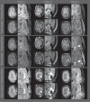
Deep learning-based Intraoperative MRI reconstruction
- PMID: 39998750
- PMCID: PMC11861787
- DOI: 10.1186/s41747-024-00548-9
Deep learning-based Intraoperative MRI reconstruction
Abstract
Background: We retrospectively evaluated the quality of deep learning (DL) reconstructions of on-scanner accelerated intraoperative MRI (iMRI) during respective brain tumor surgery.
Methods: Accelerated iMRI was performed using dual surface coils positioned around the area of resection. A DL model was trained on the fastMRI neuro dataset to mimic the data from the iMRI protocol. The evaluation was performed on imaging material from 40 patients imaged from Nov 1, 2021, to June 1, 2023, who underwent iMRI during tumor resection surgery. A comparative analysis was conducted between the conventional compressed sense (CS) method and the trained DL reconstruction method. Blinded evaluation of multiple image quality metrics was performed by two neuroradiologists and one neurosurgeon using a 1-to-5 Likert scale (1, nondiagnostic; 2, poor; 3, acceptable; 4, good; and 5, excellent), and the favored reconstruction variant.
Results: The DL reconstruction was strongly favored or favored over the CS reconstruction for 33/40, 39/40, and 8/40 of cases for readers 1, 2, and 3, respectively. For the evaluation metrics, the DL reconstructions had a higher score than their respective CS counterparts for 72%, 72%, and 14% of the cases for readers 1, 2, and 3, respectively. Still, the DL reconstructions exhibited shortcomings such as a striping artifact and reduced signal.
Conclusion: DL shows promise in allowing for high-quality reconstructions of iMRI. The neuroradiologists noted an improvement in the perceived spatial resolution, signal-to-noise ratio, diagnostic confidence, diagnostic conspicuity, and spatial resolution compared to CS, while the neurosurgeon preferred the CS reconstructions across all metrics.
Relevance statement: DL shows promise to allow for high-quality reconstructions of iMRI, however, due to the challenging setting of iMRI, further optimization is needed.
Key points: iMRI is a surgical tool with a challenging image setting. DL allowed for high-quality reconstructions of iMRI. Additional optimization is needed due to the challenging intraoperative setting.
Keywords: Artifacts; Brain neoplasms; Deep learning; Magnetic resonance imaging; Neurosurgical procedures.
© 2025. The Author(s).
Conflict of interest statement
Declarations. Ethics approval and consent to participate: This retrospective study was approved by the Regional Medical Ethics Committee for Oslo University Hospital (REK 367336), and informed consent was acquired prior to surgery according to a broad research approval and biobank (REK 2016/17091). Only patients over 18 years were included. Consent for publication: Not applicable. Competing interests: MWAC is a shareholder of Nico-lab International Ltd. AB is a shareholder in NordicNeuroLab AS, Bergen, Norway. The remaining authors declare no competing interests.
Figures


References
-
- Rogers CM, Jones PS, Weinberg JS (2021) Intraoperative MRI for brain tumors. J Neurooncol 151:479–490. 10.1007/s11060-020-03667-6 - PubMed
-
- Senft C, Bink A, Franz K et al (2011) Intraoperative MRI guidance and extent of resection in glioma surgery: a randomised, controlled trial. Lancet Oncol 12:997–1003. 10.1016/S1470-2045(11)70196-6 - PubMed
-
- Kubben PL, ter Meulen KJ, Schijns OEMG et al (2011) Intraoperative MRI-guided resection of glioblastoma multiforme: a systematic review. Lancet Oncol 12:1062–1070. 10.1016/S1470-2045(11)70130-9 - PubMed
-
- Pruessmann KP, Weiger M, Scheidegger MB, Boesiger P (1999) SENSE: sensitivity encoding for fast MRI. Magn Reson Med 42:952–962 - PubMed
-
- Griswold MA, Jakob PM, Heidemann RM et al (2002) Generalized autocalibrating partially parallel acquisitions (GRAPPA). Magn Reson Med 47:1202–1210. 10.1002/MRM.10171 - PubMed
MeSH terms
Grants and funding
LinkOut - more resources
Full Text Sources
Medical
