. 2025 Feb 25;20(2):e0318754.
doi: 10.1371/journal.pone.0318754. eCollection 2025.

Soluble RAGE enhances muscle regeneration after cryoinjury in aged and diseased mice

Affiliations

Soluble RAGE enhances muscle regeneration after cryoinjury in aged and diseased mice

Naftali Horwitz et al. PLoS One. .

Abstract

The Receptor for Advanced Glycation End Products (RAGE), classically considered a mediator of acute and chronic inflammatory responses, has recently been implicated by genetic knockout studies as a regulator of skeletal muscle physiology during development and following acute injury. Yet, the role of its soluble isoform, soluble RAGE (sRAGE), in muscle regeneration remains relatively unexplored. To address this knowledge gap, Adeno-Associated Virus (AAV) mediated and genetic knockin supplementation strategies were developed to specifically assess the effects of changing levels of sRAGE on muscle regeneration. We evaluated general muscle physiology and histology, including central nucleation, and myofiber size. We found that acute induction of sRAGE in aged and atherosclerotic animals accelerates muscle repair after cryoinjury. Similarly, genetic modification of the endogenous Ager gene locus to favor production of sRAGE over transmembrane RAGE accelerates repair of cryo-damaged skeletal muscle. However, increasing sRAGE via AAV delivery or using our transgenic mouse lines had no impact on muscle repair in aged or diseased mice after barium chloride (BaCl2) injury. Together, these studies identify a unique muscle regulatory activity of sRAGE that is variable across injury models and may be targeted in a context-specific manner to alter the skeletal muscle microenvironment and boost muscle regenerative output.


Conflict of interest statement

A.J.W. is a scientific advisor for Kate Therapeutics and Frequency Therapeutics, as well as a co-founder and scientific advisory board member and holds private equity in Elevian, Inc., a company that aims to develop medicines to restore regenerative capacity. Elevian also has provided sponsored research to the Wagers lab.

Figures

Fig 1
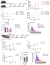
Fig 1. sRAGE accelerates regeneration in aged skeletal muscle after cryoinjury.
A) ELISA measurement of serum sRAGE concentration in 4, 7, 12 and 18 month old C57BL/6J mice (n =  5 male mice per condition). B) Design of AAV9-sRAGE viral vector delivery system comprising of a truncated constitutive CBA hybrid (CBh) promoter, an miRNA-122 target site – to decrease liver targeted expression – alongside the sRAGE transgene, all packaged in a self-complementary backbone. C) Experimental scheme for muscle regeneration assay in vehicle (FBB) and AAV9-sRAGE injected Adult (7–8 months old), and aged (18 months old) mice. D) In vivo kinetics of serum sRAGE levels in vehicle (FBB) and AAV9-sRAGE injected adult mice (n ≥  7 male mice per condition). Enumeration of the mean CSA (E) and distribution (F) of regenerating (centrally nucleated) muscle fibers in vehicle (FBB) and sRAGE treated, adult mice at 7 days after cryoinjury (n ≥  7 male mice per condition). G) In vivo kinetics of serum sRAGE levels in vehicle (FFB) and AAV9-sRAGE injected aged mice (n =  10 male mice per condition). H & I) Enumeration of the mean CSA (H) and distribution (I) of regenerating (centrally nucleated) muscle fibers in vehicle (FFB) and sRAGE treated, aged mice at 7 days post cryoinjury (n =  10 male mice per condition). Quantification of body weight (J), grid hang time (K), exercise endurance (L), and serum glucose levels (M) in vehicle (FFB) and sRAGE treated, aged (18 months old) mice. (n =  10 male mice per condition). Dots represent data for individual animals overlaid with mean ±  SD. Data analyzed for statistical significance by one-way ANOVA with Tukey post hoc test (A, D, G) or Student’s two-tailed unpaired t test (E, H, J, K, L, M). Myofiber size distributions analyzed by Mann-Whitney U test (F, I).
Fig 2
Fig 2. sRAGE enhances skeletal muscle regeneration after cryo-damage in a mouse model of atherosclerosis.
Experimental scheme for muscle regeneration assay in vehicle (FBB) and AAV9-sRAGE injected diabetic (Leprdb) and wild-type (WT) mice. B) In vivo kinetics of serum sRAGE levels in AAV9-sRAGE or vehicle (FFB) injected diabetic (Leprdb) and wild-type (WT) mice (n =  6 male mice per condition). C) Body weight of diabetic (Leprdb) and wild-type (WT) animals following vehicle (FFB) or AAV9-sRAGE administration (n =  6 male mice per condition). D) Serum glucose levels of diabetic (Leprdb) and wild-type (WT) animals following vehicle (FFB) or AAV9-sRAGE administration (n =  6 male mice per condition). Black brackets are shown for those comparisons that are statistically significant. Enumeration of the mean CSA (E) and distribution (F) of regenerating (centrally nucleated) muscle fibers in vehicle (FFB) and AAV9-sRAGE treated, diabetic (Leprdb) and wild-type (WT) mice at 7 days after cryoinjury (n =  6 male mice per condition). G) Experimental scheme for muscle regeneration assay in vehicle (FBB) and AAV9-sRAGE injected atherosclerotic (ApoE-null) and wild-type (WT) mice. H) In vivo kinetics of serum sRAGE levels in vehicle (FFB) and AAV9-sRAGE injected atherosclerotic (ApoE-null) animals (n =  6 male mice per condition). Body weight (I) and serum glucose levels (J) of atherosclerotic (ApoE-null) mice following vehicle (FFB) and AAV9-sRAGE administration (n =  6 male mice). Enumeration of the mean CSA (K) and distribution (L) of regenerating (centrally nucleated) muscle fibers in vehicle (FFB) and AAV9-sRAGE treated, atherosclerotic (ApoE-null) mice at 7 days after cryoinjury (n =  6 male mice per condition). Dots represent data for individual animals overlaid with mean ±  SD. Data analyzed for statistical significance by two-way ANOVA with Tukey post hoc test (B, C, D), one way ANOVA with Tukey post hoc test (E, H, I, J) or Student’s two-tailed unpaired t test (K). Myofiber size distributions analyzed by Mann-Whitney U test and Kruskal-Wallis test (F, L). All mice were 2–3 months of age at the time of study entry.
Fig 3
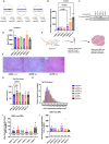
Fig 3. Endogenous and lifelong expression of a transgene encoding sRAGE enhances skeletal muscle regeneration post cryoinjury.
A) Genetic composition at the AGER locus of transgenic and wild type mice used in this study. AGER KO animals were previously generated via a cre-mediated loxP recombination of essential elements (exons 2 to 7) of the murine RAGE gene. sRAGE transgenic animals were previously generated by replacing exons 10 − 11 of the Ager gene, which encode the transmembrane and cytosolic signaling domains of the murine RAGE, with a bicistronic control element 2A-linked EGFP expression cassette. Therefore, this animal is a full-length RAGE-null and the resulting sRAGE transgene is expressed in RAGE-expressing cell types using its normal gene regulatory elements. B) ELISA measurement of serum sRAGE concentration in mice of the indicated genotypes (n ≥  5 male mice per condition). C) Confirmation of C-terminal RAGE expression in mice bearing at least one Ager + allele, and absence in mice bearing only Agers or Ager- alleles, via Western blot. D) Enumeration of the mean CSA of muscle fibers in uninjured mice of the indicated genotypes (n = 3 male mice per condition). E) Experimental scheme for muscle regeneration assay in RAGE knockout and transgenic mice. F) Representative H&E stained images of regenerating muscle from Ager + / + , Agers/ + and Agers/s mice subjected 7 days previously to cryoinjury. Enumeration of the mean CSA (G) and distribution (H) of regenerating (centrally nucleated) muscle fibers in wild-type, knockout and transgenic mice at 7 days after cryoinjury (n ≥  3 male mice per condition). Quantification of engrafted dystrophin + muscle fibers following transplantation of 3000 (I) or 6000 (J) Sca1-; Cd45-; Cd11b-, Ter119-, Cd31-; ItgB7 + ; CXCR4 + MuSCs from the indicated genotypes of mice (wild-type, knockout or transgenic) into the pre-injured muscles of mdx recipient mice (n ≥  5 male mice per condition). Dots represent data for individual animals overlaid with mean ±  SD. Data analyzed for statistical significance by one-way ANOVA with Tukey post hoc test (B, D, G, I, J). Myofiber size distributions analyzed by Kruskal-Wallis test (H). All mice were 7–8 months of age at the time of study entry.

References

    1. Nguyen M-H, Cheng M, Koh TJ. Impaired muscle regeneration in ob/ob and db/db mice. Sci World J. 2011;11:1525–35. doi: 10.1100/tsw.2011.137 - DOI - PMC - PubMed
    1. Kang J, Albadawi H, Patel VI, Abbruzzese TA, Yoo J-H, Austen WG Jr, et al.. Apolipoprotein E-/- mice have delayed skeletal muscle healing after hind limb ischemia-reperfusion. J Vasc Surg. 2008;48(3):701–8. doi: 10.1016/j.jvs.2008.04.006 - DOI - PubMed
    1. Jang YC, Sinha M, Cerletti M, Dall’Osso C, Wagers AJ. Skeletal muscle stem cells: effects of aging and metabolism on muscle regenerative function. Cold Spring Harb Symp Quant Biol. 2011;76:101–11. doi: 10.1101/sqb.2011.76.010652 - DOI - PubMed
    1. Espino-Gonzalez E, Dalbram E, Mounier R, Gondin J, Farup J, Jessen N, et al.. Impaired skeletal muscle regeneration in diabetes: from cellular and molecular mechanisms to novel treatments. Cell Metab. 2024;36(6):1204–36. doi: 10.1016/j.cmet.2024.02.014 - DOI - PubMed
    1. Dong H, Zhang Y, Huang Y, Deng H. Pathophysiology of RAGE in inflammatory diseases. Front Immunol. 2022;13:931473. doi: 10.3389/fimmu.2022.931473 - DOI - PMC - PubMed

Substances

LinkOut - more resources