. 2024 Oct 11;10(41):eadq6795.
doi: 10.1126/sciadv.adq6795. Epub 2024 Oct 11.

CLIC5 promotes myoblast differentiation and skeletal muscle regeneration via the BGN-mediated canonical Wnt/β-catenin signaling pathway

Affiliations

CLIC5 promotes myoblast differentiation and skeletal muscle regeneration via the BGN-mediated canonical Wnt/β-catenin signaling pathway

Xin Zhang et al. Sci Adv. .

Abstract

Myoblast differentiation plays a vital role in skeletal muscle regeneration. However, the protein-coding genes controlling this process remain incompletely understood. Here, we showed that chloride intracellular channel 5 (CLIC5) exerts a critical role in mediating myogenesis and skeletal muscle regeneration. Deletion of CLIC5 in skeletal muscle leads to reduced muscle weight and decreases the number and differentiation potential of satellite cells. In vitro, CLIC5 consistently inhibits myoblast proliferation while promoting myotube formation. CLIC5 promotes myogenic differentiation by activating the canonical Wnt/β-catenin signaling pathway in a biglycan (BGN)-dependent manner. CLIC5 deletion impairs muscle regeneration. Paired box gene 7 (Pax7) expression and the activity of BGN-mediated canonical Wnt/β-catenin signaling are reduced in CLIC5-deficient mice. Conversely, increasing CLIC5 levels in skeletal muscles enhances muscle regeneration capacity. In conclusion, our findings underscore CLIC5 as a pivotal regulator of myogenesis and skeletal muscle regeneration, functioning through interaction with BGN to activate the canonical Wnt/β-catenin signaling pathway.


Figures

Fig. 1.
Fig. 1.. Loss of CLIC5 reduces muscle weight and decreases the number and differentiation potential of satellite cells.
Body weight of (A) male and (B) female mice from the age of 1 to 9 weeks. Muscle mass of (C) TA and (D) GAS in adult WT and CLIC5MKO mice. (E) Flow cytometry analysis of CD31, CD45, Sca1, and α7 integrin expression in whole musclve–derived cells. APC, allophycocyanin; FITC, fluorescein isothiocyanate; FSC-H, forward scatter height; FSH-W, forward scatter width; P2, CD31- cell populations. (F) The percentage of satellite cells in whole muscle–derived cells revealed by flow cytometry analysis. (G and H) Immunofluorescence of Pax7 and Laminin and satellite cells per area of TA muscle cross sections. Magnification, ×60. (I) Immunofluorescence analysis for myosin in satellite cells cultured for 3 days in DM. Magnification, ×10. (J) Differentiation index and fusion index. Differentiation index is calculated as the percentage of nuclei in myosin+ cells. Fusion index is calculated as the percentage of nuclei in myosin+ myotubes. Structures containing at least two nuclei are considered as a myotube. (K) Relative mRNA levels of Myf5, MyoD, MyoG, and Myomaker and (L and M) Western blot analysis of MyoG in satellite cells cultured in DM for 1, 2, or 3 days (D1, D2, or D3). GAPDH, glyceraldehyde-3-phosphate dehydrogenase. n = 16 to 20 for (A) to (D) and 3 for (E) to (M). The data present means ± SEM. *P < 0.05 and **P < 0.01.
Fig. 2.
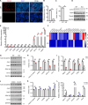
Fig. 2.. CLIC5 mediates the balance of proliferation and differentiation of myoblasts.
(A and B) Representative photographs of EdU staining in control (siCtrl) and CLIC5 knockdown (siCLIC5) C2C12 myoblasts. Nuclei were stained with Hoechst. Magnification, ×10. (C) Cell cycle analysis of siCtrl and siCLIC5 C2C12 myoblasts via flow cytometry. (D) Expression analysis of cell cycle–related genes in siCtrl and siCLIC5 myoblasts cultured in growth medium. (E) Immunofluorescence analysis for myosin in siCtrl and siCLIC5 C2C12 myoblasts after 4 days in DM. Magnification, ×10. (F) Differentiation index calculated as the percentage of nuclei in myosin+ cells and fusion index calculated as the percentage of nuclei in myosin+ myotubes. Structures containing at least two nuclei are considered as a myotube. (G) Western blot analysis of CLIC5, MyoG, and myosin in siCtrl and siCLIC5 C2C12 myoblasts after 4 days in DM. (H and I) Representative photographs of EdU staining in control (vector) and CLIC5-overexpressing C2C12 myoblasts. Nuclei were stained with Hoechst. Magnification, ×10. (J) Cell cycle analysis of control and CLIC5-overexpressing C2C12 myoblasts. (K) Expression analysis of cell cycle–related genes in control and CLIC5-overexpressing C2C12 myoblasts cultured in growth medium. (L) Immunofluorescence analysis for myosin in control and CLIC5-overexpressing C2C12 myoblasts after 4 days in DM. Magnification, ×10. (M) Differentiation index and fusion index. (N) Western blot analysis of Flag, MyoG, and myosin in control and CLIC5-overexpressing C2C12 myoblasts after 4 days in DM. The data present means ± SEM (n = 3). *P < 0.05 and **P < 0.01.
Fig. 3.
Fig. 3.. CLIC5 promotes myoblast differentiation and activates the canonical Wnt/β-catenin signaling pathway.
(A) Immunofluorescence analysis for myosin in WT and CLIC5-KO C2C12 myoblasts after 4 days in DM. Magnification, ×10. (B) Differentiation index. (C) Fusion index. (D) Western blot analysis of MyoG and myosin in WT and KO C2C12 myoblasts after 4 days in DM. (E) The mRNA expression levels of selected genes in WT and KO C2C12 myoblasts by RNA-seq. (F) Heatmap of DEGs enriched in Wnt signaling pathway in WT and KO C2C12 myoblasts. (G and H) Western blot analysis of active β-catenin, Axin1, Axin2, GSK-3β, and CK1α in WT and KO C2C12 myoblasts cultured in growth medium. (I) Relative mRNA levels of Wnt-targeted genes expressed in WT and KO C2C12 myoblasts. (J and K) Western blot analysis of active β-catenin, Axin1, Axin2, GSK-3β, and CK1α in control and CLIC5-overexpressing C2C12 myoblasts cultured in growth medium. (L) Relative mRNA levels of Wnt-targeted genes expressed in control and CLIC5-overexpressing C2C12 myoblasts. The data present means ± SEM (n = 3). *P < 0.05 and **P < 0.01.
Fig. 4.
Fig. 4.. CLIC5 interacts with BGN and promotes its expression.
(A) Immunofluorescence staining of BGN in C2C12 myoblasts. Magnification, ×20. (B) Western blot analysis of BGN in cytosol and nuclear fractions of C2C12 myoblasts. n = 3. (C) Immunofluorescence staining of CLIC5 and BGN in C2C12 myoblasts. Magnification, ×40. (D) Reciprocal coimmunoprecipitation (IP) analysis between Flag-tagged CLIC5 and Myc-tagged BGN in HEK293T cells. IB, immunoblotting. (E) CLIC5/BGN binding domain identification. Full-length and C-terminal truncated CLIC5 were overexpressed with HA-tagged BGN in HEK293T cells. Immunoblot for HA after anti–green fluorescent protein (GFP) coimmunoprecipitation. (F) Immunofluorescence staining of BGN in WT and CLIC5-KO C2C12 myoblasts. Magnification, ×60. (G to H) Western blot analysis of BGN in cytosol and plasma membrane (PM) fractions of WT and KO C2C12 myoblasts. (I) Western blot analysis of BGN after CLIC5 overexpression in C2C12 myoblasts. The data present means ± SEM (n = 3). **P < 0.01.
Fig. 5.
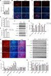
Fig. 5.. CLIC5 promotes myoblast differentiation and activates canonical Wnt/β-catenin signaling pathway through BGN.
(A) Western blot analysis of BGN in C2C12 myoblasts cultured in growth medium (D0) or in DM for 2, 4, or 6 days. Immunofluorescence analysis for myosin in siCtrl and siBGN C2C12 myoblasts (B) or myoblasts stably transfected with CLIC5 plasmid (C) after 4 days in DM. Magnification, ×10. (D) Differentiation index. (E) Fusion index. (F and G) Western blot analysis of active β-catenin, Axin1, Axin2, GSK-3β, and CK1α in siCtrl and siBGN C2C12 myoblasts stably transfected with CLIC5 plasmid. Immunofluorescence analysis for myosin (H), Western blot analysis of CLIC5, BGN, and canonical Wnt/β-catenin signaling pathway (I and J), as well as relative mRNA levels of Wnt-targeted genes (K) in siCtrl, siCLIC5, and siCLIC5 C2C12 myoblasts transfected with BGN plasmid after 6 days in DM. Magnification, ×10. The data present means ± SEM (n = 3). *P < 0.05 and **P < 0.01.
Fig. 6.
Fig. 6.. Loss of CLIC5 impairs skeletal muscle regeneration.
(A) Schematic outline of cardiotoxin (CTX) injection and sample collection. (B) Hematoxylin and eosin (H&E) staining of injured TA muscle in WT and CLIC5MKO mice at days 0, 3, 7 and 14 after injury. Magnification, ×20. (C) Immunofluorescence analysis of eMyHC+ fibers in TA muscles at days 3, 7, and 14 after injury. Scale bar, 50 μm. (D and E) Western blot analysis of CLIC5, MyoG, Pax7, BGN, active β-catenin, Axin2, GSK-3β, and CK1α in the TA muscle of WT and CLIC5MKO mice at day 3 after injury. The data present means ± SEM (n = 3). *P < 0.05 and **P < 0.01.
Fig. 7.
Fig. 7.. Increasing CLIC5 in the skeletal muscle enhances its regeneration capacity.
(A) Schematic outline of the administration of AAV9:empty vector or AAV9:CLIC5 vectors, CTX injection, and sample collection. (B) H&E staining of injured TA muscle at days 0, 3, 7, and 14 after injury. Magnification, ×20. (C) Immunofluorescence analysis of eMyHC+ fibers in TA muscles at days 3, 7, and 14 after injury. Scale bar, 50 μm. (D and E) Western blot analysis of CLIC5, MyoG, Pax7, BGN, active β-catenin, Axin2, GSK-3β, and CK1α in the TA muscle of mice administered AAV9:empty vector or AAV9:CLIC5 vectors at day 3 after injury. The data present means ± SEM (n = 5). *P < 0.05 and **P < 0.01.

References

    1. Wosczyna M. N., Rando T. A., A muscle stem cell support group: Coordinated cellular responses in muscle regeneration. Dev. Cell 46, 135–143 (2018). - PMC - PubMed
    1. Janssen I., Heymsfield S. B., Wang Z. M., Ross R., Skeletal muscle mass and distribution in 468 men and women aged 18–88 yr. J. Appl. Physiol. 89, 81–88 (2000). - PubMed
    1. Yin H., Price F., Rudnicki M. A., Satellite cells and the muscle stem cell niche. Physiol. Rev. 93, 23–67 (2013). - PMC - PubMed
    1. Chargé S. B., Rudnicki M. A., Cellular and molecular regulation of muscle regeneration. Physiol. Rev. 84, 209–238 (2004). - PubMed
    1. Zanou N., Gailly P., Skeletal muscle hypertrophy and regeneration: Interplay between the myogenic regulatory factors (MRFs) and insulin-like growth factors (IGFs) pathways. Cell. Mol. Life Sci. 70, 4117–4130 (2013). - PMC - PubMed

MeSH terms

LinkOut - more resources