. 2025 Feb 25;16(1):1973.
doi: 10.1038/s41467-025-57204-y.

Structural basis for lipid-mediated activation of G protein-coupled receptor GPR55

Affiliations

Structural basis for lipid-mediated activation of G protein-coupled receptor GPR55

Tobias Claff et al. Nat Commun. .

Abstract

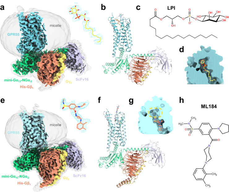
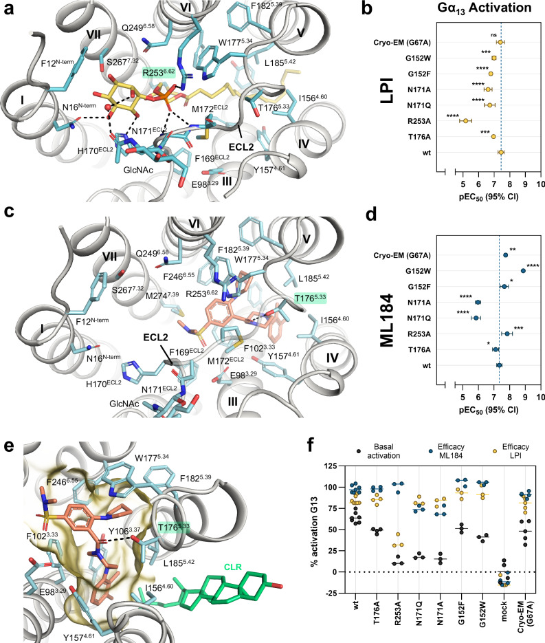
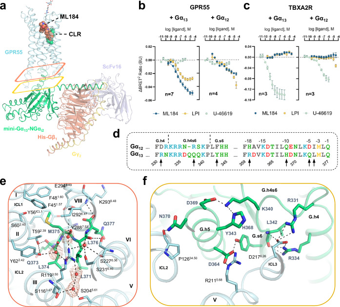
GPR55 is an orphan G protein-coupled receptor (GPCR) and represents a promising drug target for cancer, inflammation, and metabolic diseases. The endogenous activation of lipid GPCRs can be solely mediated by membrane components and different lipids have been proposed as endogenous activators of GPR55, such as cannabinoids and lysophosphatidylinositols. Here, we determine high-resolution cryo-electron microscopy structures of the activated GPR55 in complex with heterotrimeric G13 and two structurally diverse ligands: the putative endogenous agonist 1-palmitoyl-2-lysophosphatidylinositol (LPI) and the synthetic agonist ML184. These results reveal insights into ligand recognition at GPR55, G protein coupling and receptor activation. Notably, an orthosteric binding site opening towards the membrane is observed in both structures, enabling direct interaction of the agonists with membrane lipids. The structural observations are supported by mutagenesis and functional experiments employing G protein dissociation assays. These findings will be of importance for the structure-based development of drugs targeting GPR55.


Conflict of interest statement

Competing interests: All authors are employees of Boehringer Ingelheim Pharma GmbH & Co. KG.

Figures

Fig. 1
Fig. 1. Architecture of the GPR55-G13 signaling complex with lipid and synthetic agonists.
a Cryo-EM map (consensus, EMDB-51285) of the GPR55-Gα13β1γ2-ScFv16-LPI complex at two different contour levels. The enlarged cryo-EM map for LPI (yellow sticks) is shown in blue mesh. b Full model corresponding to the signaling complex of (a) (shown as cartoon representation). c Chemical structure of LPI. d Overview of ligand binding pocket position of LPI. e Cryo-EM map (consensus, EMDB-51281) of the GPR55-Gα13β1γ2-ScFv16-ML184 complex at two different contour levels. The enlarged cryo-EM map for ML184 (salmon sicks) is shown in blue mesh. f Full model corresponding to the signaling complex of (d). g Overview of ligand binding pocket position of ML184. The yellow circle highlights the position of the polar head group of LPI. h Chemical structure of ML184.
Fig. 2
Fig. 2. Agonist recognition at GPR55.
a Ligand binding pocket of LPI (yellow sticks). The amino acid side chain and backbone that show interactions with LPI are shown as cyan sticks. Hydrogen bonds are indicated as black dashed lines. b Effect of GPR55 mutants on LPI potency as determined by G protein dissociation assays with Gα13. Data represent means ± 95% confidence interval (CI) from 3–7 independent experiments as indicated in Supplementary Table S3. c ligand binding pocket of ML184. d Effect of GPR55 mutants on ML184 potency as determined by G protein dissociation assays with Gα13. Data represent means ± 95% CI from 3–7 independent experiments as indicated in Supplementary Table S3. e Magnified view of the ML184 (salmon sticks) binding pocket (as in a). The binding pocket surface was displayed in yellow with the ECL2 surface hidden for better visibility of the binding pocket. f Efficacy and constitutive activity at different GPR55 mutants. The degree of activation was calculated by normalization of BRET² ratios (BRET² ratio of 30 µM ligand for efficacy or basal BRET² ratios of 1.5% DMSO) to the respective BRET² ratios for the wt GPR55 plus G13-biosensor at 30 µM ML184 (100% activation) and for a mock-transfection plus G13-biosensor (0% activation). The pEC50 means of all seven mutations were compared with the pEC50 mean of the wt GPR55 to evaluate statistically significant differences using ordinary one-way ANOVA with Dunnett’s post-hoc test (adjusted P-values: *p < 0.05, **p < 0.01, ***p < 0.001, ****p < 0.0001, for absolute P-values see Supplementary Table S3).
Fig. 3
Fig. 3. Membrane opening close to orthosteric pocket and cholesterol binding site.
a Side view of the GPR55-LPI complex (cyan surface representation and yellow spheres, respectively) shows membrane gate into the hydrophobic channel of the orthosteric binding pocket between helices IV and V. Membrane lipids were not resolved in this structure. b Side view of the GPR55-ML184 (light cyan surface representation) complex resolved a CLR molecule (green spheres) bound in a similar cleft between helices IV and V. c Superimposition of the GPR55-LPI and GPR55-ML184 structures (receptor in cartoon representation, with side chains that contact CLR as sticks) show the CLR (green sticks) binding pocket with direct contacts to ML184 (salmon sticks). Subtle sidechain rearrangements in the LPI structure within the CLR interface are observed. The dotted yellow line represents the distance measurement (in Å) between K1805.37 and CLR.
Fig. 4
Fig. 4. Molecular dynamics simulations investigate the CLR binding pocket stability.
a, b Averaged CLR mass density plot generated from martini coarse-grained simulations for (a) martini system 1 (CLR is placed in the structure pose) and (b) for martini system 2 (CLR was removed), the yellow circle highlights the CLR density in both figures. c Superimposition of final structures from all-atom system 1 (shown in cyan) containing CLR and ML184 with system 4 (shown in yellow) where ML184 and CLR were removed shows that significant changes in the protein conformation is observed (black arrow) when the CLR molecule was removed from the binding site. The structural CLR molecule is shown in green and ML184 is shown in orange. d Enlarged view of the CLR binding pocket and (e) surface representation show that CLR binding site is occluded significantly. The all-atom simulation without ML184 and CLR also has an impact on the ligand binding pocket as shown in (f, g). h Overview of four simulated all-atom systems with the RMSD of the ML184 binding pocket calculated from the representative structure of the most populated cluster, compared to system 1. n.a.: not applicable.
Fig. 5
Fig. 5. Gα13 activation by GPR55 and selectivity over Gα12.
a Overview of the GPR55-G13-ML184 signaling complex. ML184 and CLR are shown as salmon and green spheres, respectively. Colored rectangles highlight specific G protein interaction sections as described in panels (e, f), respectively. The map quality of one water molecule deviates significantly from four well-resolved water molecules. The modeling of this water molecule was guided by its coordination with R1193.50, helix VI, and helix VII. b, c G protein dissociation assays of (b) GPR55 and (c) TBXA2R with Gα12 and Gα13. Assays were performed in HEK293H cells transiently transfected with wt Gβ3, Gγ9-GFP, and Gα12-Rluc8 or Gα13-Rluc8. Data represents means ± SEM from 3–7 independent experiments as specified in Supplementary Table S3. The negative control data for U-46619 at GPR55 and ML184 or LPI at the TBXA2R was analyzed from three independent experiments. d Sequence alignment of different G protein segments of Gα13 involved in GPR55 binding with the respective residues of Gα12. Black arrows highlight amino acid differences between Gα12 and Gα13 within 4 Å of GPR55. Residues R360 and Q338 are predominantly solvent-exposed and not shown in the following panels. e Protein-protein interface between Gα13 (green cartoon and sticks) and GPR55 (cyan cartoon and sticks) with a focus on the C-terminal α5-helix of Gα13. The cryo-EM composite map (EMDB-51284) for five water molecules is shown with orange mesh. f Protein-protein interactions of residues between the α4-helix and β6-sheet of Gα13 as well as of the initial α5-helix residues with GPR55.

Similar articles

Cited by

References

    1. Hauser, A. S., Attwood, M. M., Rask-Andersen, M., Schiöth, H. B. & Gloriam, D. E. Trends in GPCR drug discovery: new agents, targets and indications. Nat. Rev. Drug Discov.16, 829–842 (2017). - PMC - PubMed
    1. Jacobson, K. A. New paradigms in GPCR drug discovery. Biochem. Pharmacol.98, 541–555 (2015). - PMC - PubMed
    1. Lee, A. G. How lipids affect the activities of integral membrane proteins. Biochim. Biophys. Acta Biomembr.1666, 62–87 (2004). - PubMed
    1. Mizuno, H. & Kihara, Y. Druggable lipid signaling pathways. Adv. Exp. Med. Biol.1274, 223–258 (2020). - PubMed
    1. Smith, N. J. & Murray, F. Shifting our perspective on orphan G protein-coupled receptors. Nat. Struct. Mol. Biol.31, 582–583 (2024). - PubMed

LinkOut - more resources