. 2025 Feb 25;15(1):6762.
doi: 10.1038/s41598-025-88037-w.

Histomorphometric analysis of anterior cruciate ligament bundles and anatomical insights into injury mechanisms

Affiliations

Histomorphometric analysis of anterior cruciate ligament bundles and anatomical insights into injury mechanisms

In-Seung Yeo et al. Sci Rep. .

Abstract

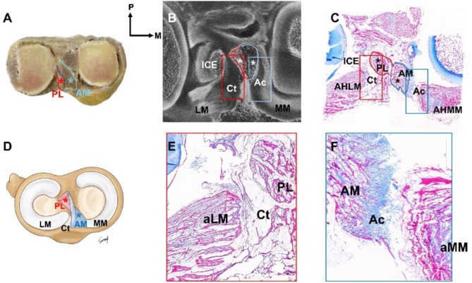
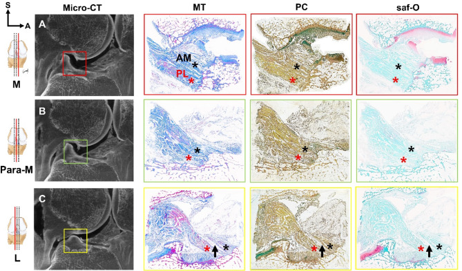
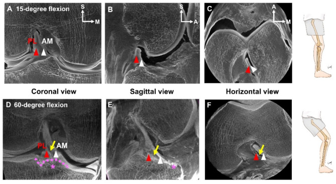
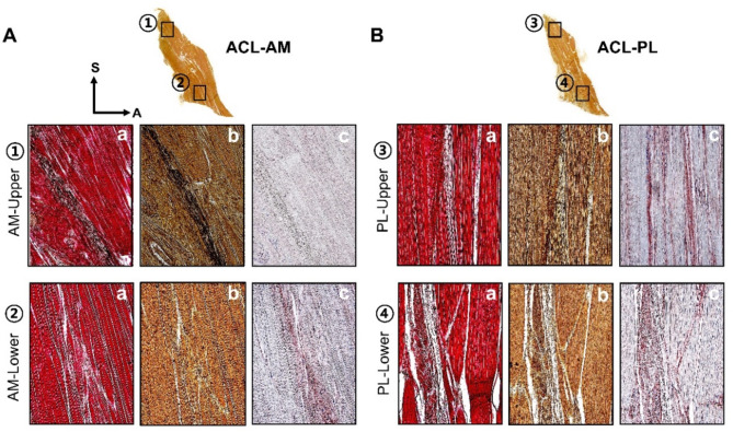
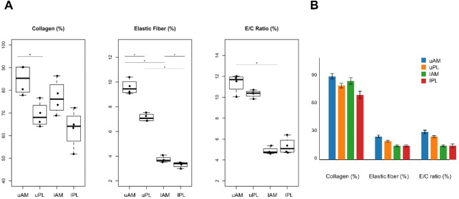
The anterior cruciate ligament consists of two bundles, the anteromedial and posterolateral bundles, which are frequently associated with meniscal dysfunction. Despite previous studies investigating the relationship between biomechanical instability and injury, a comprehensive histological analysis of the anatomical aspects contributing to injury and degenerative changes and the structural connectivity between the anterior cruciate ligament and the meniscus is lacking. Masson's trichrome, pentachrome, Safranin-O, and modified Verhoeff-Van Gieson histological stains and microcomputed tomography were used in this analysis. The anteromedial bundle of the anterior cruciate ligament is tightly connected to the medial meniscus via articular cartilage, whereas the posterolateral bundle is loosely connected to the transition zone of the lateral meniscus via connective tissue. Due to the differences in the structural connectivity between the meniscus and each anterior cruciate ligament bundle, the degree of deformation of the space between the two bundles varies significantly with knee flexion angle. Furthermore, the two bundles exhibit histological differences in the ratio of elastic fibers to collagen at regions. Specifically, the ratios of the upper and lower parts were 11.36 ± 0.90% and 4.87 ± 0.34%, respectively, for the anteromedial bundle, and 10.33 ± 0.37% and 5.32 ± 0.78%, respectively, for the posterolateral bundle.

Keywords: Anatomy; Anterior cruciate ligament; Articular cartilage; Lateral meniscus; Medial meniscus.


Conflict of interest statement

Declarations. Competing interests: The authors declare no competing interests.+ After this sentences, there is missing information about the Funding. Could you please add it as it was in the original manuscript?

Figures

Fig. 1
Fig. 1
The overall morphological transverse section of the knee joint complex (AF). These structures were subsequently compared and analyzed using microcomputed tomography ((micro-CT, (B). Applying modified Masson’s trichrome staining (MT) confirmed the connectivity of the anteromedial (AM) bundle of the anterior cruciate ligament (ACL) (C; blue asterisk), posterolateral (PL) bundle of the ACL (C); red asterisk)), and meniscus (C). The AM bundle tightly connected to the anterior horn of the medial meniscus (aMM) and articular cartilage (F); Ac, blue box)), while the PL bundle loosely connected to the anterior horn of the lateral meniscus (aLM) and connective tissue (E); Ct, red box)). AM anterior cruciate ligament-anteromedial bundle, Ac articular cartilage, aLM anterior horn of lateral meniscus, aMM anterior horn of medial meniscus, Ct connective tissue, ICE intercondylar eminence, LM lateral meniscus, MM medial meniscus, PL anterior cruciate ligament-posterolateral bundle.
Fig. 2
Fig. 2
Analysis of immunohistochemical staining (MT, PC, and saf-O) in serial sagittal sections of the knee joint flexed at 15° (AC). Subsequently, the analysis results were compared using micro-CT. The space between the AM bundle (black asterisk) and the PL bundle (red asterisk) of the ACL was observed to gradually widened from the medial to the lateral side of the knee (indicated by the black arrow). This sequential arrangement of the ACL is illustrated from the red box to the yellow box (Supplemental Video 1). A: anterior, L lateral, M medial, Micro-CT micro-computed tomography, MT Masson’s trichrome staining, Para-M para-medial, PC pentachrome staining, saf-O safranin-O staining, S superior.
Fig. 3
Fig. 3
Comparative analysis of 15° and 60° of knee flexion using micro-CT (AF). The coronal view (A,D), sagittal view (B,E), and horizontal view (C,F) were analyzed for each angle to confirm morphological differences between the AM bundle (white arrowhead) and the PL bundle (red arrowhead) of the ACL. At 60 degrees of flexion, the gap between the AM bundle and the PL bundle gradually widened (Supplemental Video 2), becoming distinctly verified (indicated by the yellow arrow; DF). The transverse ligament is denoted by the pink dotted line (pink asterisk; D,E). A anterior, AM anterior cruciate ligament-anteromedial bundle, LM lateral meniscus, MM medial meniscus, M medial, P posterior, PL anterior cruciate ligament-posterolateral bundle, S superior.
Fig. 4
Fig. 4
Identification of histomorphological differences between the upper and lower regions of both the AM and PL bundles of the ACL using immunohistochemical methods (VG (a), PC (b), Collagen (c); (A,B). Modified Verhoeff–Van Gieson staining (a) confirmed the characteristics of collagen and elastic fibers in magnified images. Collagen is indicated in red, and black dots represent elastic fibers. In Pentachrome staining (b), elastic fibers appear as brownish collagen clusters with black dots and were further analyzed using collagen immunohistochemical staining. The upper region of the AM bundle (A) displays a dense concentration of elastic fibers, whereas the lower region exhibits a sporadic, multiskewed pattern. Similarly, in the PL bundle (B), the upper region contains more elastic fibers than in the lower region. The collagen arrangement in the AM bundle (A-c) appears to be densely disorganized, while in the PL bundle (B-c), it exhibits an organized pattern with a lower collagen proportion. ACL-AM anterior cruciate ligament-anteromedial bundle, ACL-PL anterior cruciate ligament-posterolateral bundle.
Fig. 5
Fig. 5
Graphs depicting the area ratio of elastic fibers to collagen in four regions of the ACL based on Fig. 4. The ratio for the AM bundle was highest in the upper regions, at 11.36 ± 0.90% (p < 0.05), and lowest in the lower region, at 4.87 ± 0.34% (p < 0.05), (A). Analysis of the regional composition of the PL bundle revealed that the dense area accounted for 10.33 ± 0.37% and the loose area accounted for 5.32 ± 0.78% (p < 0.05), (A). As demonstrated in Figure (B), the figures for each area were presented through the master chart. The boxplots display regional differences in the E/C ratio (elastic fiber/collagen fiber area ratio), with each box indicating median, 25th and 75th percentiles, and range values. Statistical significance (p < 0.05) is indicated by asterisks. E/C ratio, elastic fiber/collagen fiber area ratio; lAM, lower anteromedial bundle; lPL, lower posterolateral bundle; uAM, upper anteromedial bundle; uPL, upper posterolateral bundle.

Similar articles

Cited by

References

    1. Sullivan, D., Levy, I. M., Sheskier, S., Torzilli, P. A. & Warren, R. F. Medial restraints to anterior-posterior motion of the knee. J. Bone Joint Surg. Am.66, 930–936. 10.2106/00004623-198466060-00015 (1984). - PubMed
    1. Fu, F., Shen, H., Starman, W., Okeke, J. S., Irrgang, J. J. & N., & Primary anatomic double-bundle Anterior Cruciate Ligament Reconstruction: a preliminary 2-Year prospective study. Am. J. Sports Med.36, 1263–1274. 10.1177/0363546508314428 (2008). - PubMed
    1. Jerosch, J., Prymka, M. & Castro, W. H. Proprioception of knee joints with a lesion of the medial meniscus. Acta Orthop. Belg.62, 41–45 (1996). - PubMed
    1. Smith, J. P. I. I. I. & Barrett, G. R. Medial and lateral meniscal tear patterns in anterior cruciate ligament-deficient knees: a prospective analysis of 575 tears. Am. J. Sports Med.29, 415–419. 10.1177/03635465010290040501 (2001). - PubMed
    1. Wyatt, R., Inacio, W., Liddle, M. C., Maletis, G. B. & K. D., & Factors associated with meniscus repair in patients undergoing anterior cruciate ligament reconstruction. Am. J. Sports Med.41, 2766–2771. 10.1177/0363546513503287 (2013). - PubMed

MeSH terms

LinkOut - more resources