. 2025 Mar;57(3):515-521.
doi: 10.1038/s41588-025-02097-2. Epub 2025 Feb 25.

Cancer-independent somatic mutation of the wild-type NF1 allele in normal tissues in neurofibromatosis type 1

Affiliations

Cancer-independent somatic mutation of the wild-type NF1 allele in normal tissues in neurofibromatosis type 1

Thomas R W Oliver et al. Nat Genet. 2025 Mar.

Abstract

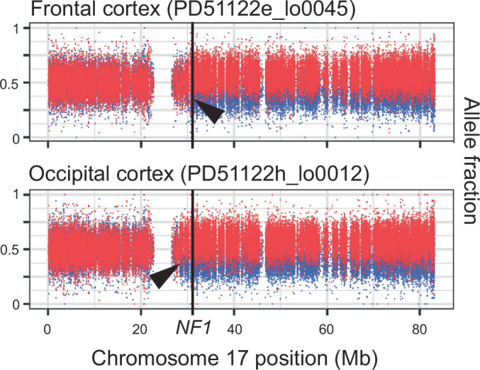
Cancer predisposition syndromes mediated by recessive cancer genes generate tumors via somatic variants (second hits) in the unaffected allele. Second hits may or may not be sufficient for neoplastic transformation. Here we performed whole-genome and whole-exome sequencing on 479 tissue biopsies from a child with neurofibromatosis type 1, a multisystem cancer-predisposing syndrome mediated by constitutive monoallelic NF1 inactivation. We identified multiple independent NF1 driver variants in histologically normal tissues, but not in 610 biopsies from two nonpredisposed children. We corroborated this finding using targeted duplex sequencing, including a further nine adults with the same syndrome. Overall, truncating NF1 mutations were under positive selection in normal tissues from individuals with neurofibromatosis type 1. We demonstrate that normal tissues in neurofibromatosis type 1 commonly harbor second hits in NF1, the extent and pattern of which may underpin the syndrome's cancer phenotype.


Conflict of interest statement

Competing interests: I.M. is a cofounder and consultant of Quotient Therapeutics. D.H. provides consultancy to AstraZeneca/MedImmune, Alexion Pharmaceuticals, Bayer, Biodexa, Roche/Genentech and Novartis, as well as expert testimony to AstraZeneca and Novartis, and his expenses are covered by Alexion Pharmaceuticals, Boehringer Ingelheim, Roche/Genentech and Novartis. The other authors declare no competing interests.

Figures

Fig. 1
Fig. 1. Concepts and experimental approach.
a, The second mutation in a recessive tumor predisposition syndrome is typically thought to lead to neoplasia. b, Some second hits may be found in the adjacent normal tissue to a childhood cancer, indicating that their presence is insufficient for neoplastic transformation. c, The possibility remains that second hits may be sustained in normal tissues that are independent of the cancer cell lineage. d, Histological images of three illustrative microdissected tissues are shown. The layers of the cerebellar cortex are annotated on the uppermost image. The light blue outlines with yellow arrowheads on the images are representative regions microdissected. Scale bars = 500, 250 and 250 µm (top to bottom). e, Experiment overview, detailing the number of bulk- and LCM-derived sequences generated per anatomical region per child. Please note that this includes all biopsies, irrespective of tumor involvement. A version of this table, limited to the tumor biopsies used in the high-grade glioma driver mutation identification, is provided as Supplementary Table 2. ML, molecular layer; PL, Purkinje layer; GL, granular layer; WM, white matter; CNS, central nervous system; PNS, peripheral nervous system; LCM, laser capture microdissection.
Fig. 2
Fig. 2. NF1 mutations and driver events identified by sequencing bulk tissues or microdissections.
Individual driver variants can be found in the lists of mutations provided in Supplementary Tables 10–14. A single asterisk indicates that the identification of the LOH event was only possible because we could phase parental alleles (Methods). The low cell fraction of many of these meant that it was not possible to determine the breakpoint. When counting LOH events, those called from sequences derived from the same original bulk biopsy are treated as the same event, and those from different biopsies are treated as unique. These events should not be considered when comparing mutations against the other two children because their SNP alleles on chromosome 17 could not be phased. Double asterisks indicate that in sequences from the high-grade glioma, we only considered mutations in genes recognized in a large meta-analysis to be drivers of these neoplasms (Methods). Triple asterisks indicate that one additional MSH6 frameshift mutation (p.F1088fs*2) was noted in the spinal cord (PD51122v_lo0008) and spleen (PD51122z_lo0017) of the child with neurofibromatosis type 1; this, however, remained heterozygous without evidence of hypermutation and did not co-occur with somatic NF1 mutation, making it of uncertain significance. Four asterisks indicate that chromosome 11p LOH was identified in a single sample after manual inspection of the copy number output, although it was too low a fraction to be detected by the copy number caller. Del, deletion; Inv, inversion.
Fig. 3
Fig. 3. NF1 null clones pervade the normal tissues of individuals with neurofibromatosis type 1.
a, Loss of the wild-type NF1 allele is an independent event in each of the three neoplasms in PD51122. The allele frequency (y axis) represents a rolling window of 50 SNPs. Gridlines are present for matching to coordinates. b, The substitution count and median VAF in normal brain, color-coded by the presence of biallelic NF1 mutation. c, Schematic representation of the brain and spinal cord outlining the location of each somatic NF1 mutation in normal tissue from PD51122 discovered by different sequencing methods (before genotyping). The VAF of each mutation is shown in the table (2sf). The figure is created with BioRender.com. d, Distribution of somatic NF1 mutations across tissues from PD51122 (above the locus-specific error rate; Methods). The number within the box indicates the number of mutated samples from that tissue (red if >0). e, Pairwise comparison of the number of substitutions shared between whole-genome sequences of normal biopsies, according to whether they possess the same NF1 second hit. The black line represents the interquartile range. The black dot is the median. P values were generated using one-sided permutation tests (Methods). ‘n’ refers to the number of pairwise comparisons, not samples. The CNS and MES groups exclude normal tissues with a second NF1 hit. f, The dN/dS ratios for truncating variants, according to germ layer and NF1 germline mutation status. The dot represents the maximum likelihood estimate, and the lines represent the 95% credible interval. When the lower bound of the credible interval is above 1 (red line), there is a statistically significant positive selection. Credible intervals falling below the boundary of the plot are terminated with slanted double lines. g, Normal tissues from adults with neurofibromatosis type 1 are grouped by tissue type and evaluated for an excess of nonsynonymous variants in NF1 and compared with the index children. Top, dN/dS ratios for truncating mutations (the dot represents the maximum likelihood estimate, and the lines represent the 95% credible interval); middle, counts of variants in NF1; bottom, total duplex coverage (Methods; Supplementary Note) over NF1 in each group. R, right; L, left; MES, mesoderm; WT, wild type.
Extended Data Fig. 1
Extended Data Fig. 1. Subcutaneous ankle lesion in the child with neurofibromatosis type 1.
a,b, Representative histological appearances of the tissue are shown. The tissue comprises adipose tissue, bands of fibrous tissue, a Pacinian corpuscle, thick nerve trunks and ganglia. c, The light blue outline with a yellow arrowhead indicates the region microdissected that yielded a biallelic loss of NF1 (PD51122ac_lo0013). It comprises bland spindled cells, embedded in fibrous and fibrillary stroma, that surround a blood vessel. The scale bars represent 1 mm, 1 mm and 250 µm, respectively.
Extended Data Fig. 2
Extended Data Fig. 2. Mosaic uniparental disomy of chromosome 11p in a nerve from case PD51123.
Left, a histological image of the slide from which the affected sample was microdissected. Scale bar represents 250 µm. Right, the coverage of chromosome 11 SNPs and their B-allele fraction (BAF). Gridlines are present for matching to coordinates. The B-allele here is the one inferred to be derived from the major allele within predicted haplotype blocks (red line). The BAF split was not detected by ascatPCA but detected on manual review and the output here is from Battenberg.
Extended Data Fig. 3
Extended Data Fig. 3. Histological images of normal tissues with independent NF1 null clones.
Where the clone was detected in a microdissected sample, the exact section and area sequenced are shown. For clones found in bulk samples, a representative image from the reference slide is provided. The microdissected and reference sections are 16 µm and 4 µm thick. PD51122g_lo0003 was taken from a section that had dried before slide scanning and microdissection, resulting in a suboptimal image. Consequently, the reference slide was used here to provide an overview of the approximate area microdissected. The black outlines on images represent the perimeter of the microdissection. PD51122b_lo0018 is a region of the molecular layer (ML), taken from the cerebellum. The scale bars for the images of microdissected samples represent 250 µm. The scale bars for PD51122b, PD51122e and PD51122f are 1 mm and for PD51122d 500 µm.
Extended Data Fig. 4
Extended Data Fig. 4. Brain MRI at diagnosis from the child with neurofibromatosis type 1.
FLAIR (top row, left and right images) and axial T2-weighted images (all other images; top row, middle and right images are rotated 90° anticlockwise) show typical focal areas of signal intensity (FASI) in the white matter and cerebellar cortex (blue boxes). The presence of tumor (red outline) makes it challenging in some areas to distinguish tumor from FASI (yellow boxes). No convincing FASI was found in the cerebral cortex or spinal cord, suggesting that no close correlation of the NF1 null clones and FASI could be made. Note that there is hyperintensity of both hippocampi (bottom row, left image).
Extended Data Fig. 5
Extended Data Fig. 5. Allele fraction plots for the normal tissues in the child with neurofibromatosis type 1 with the largest NF1 LOH events.
Each dot represents a heterozygous SNP that is phased to either the copy of chromosome 17 bearing the germline NF1 mutation (red) or the wild-type allele (blue). Black arrowheads indicate the approximate location of the breakpoint in each sample.
Extended Data Fig. 6
Extended Data Fig. 6. Loss of the germline mutated NF1 allele in bladder muscle.
Left, a histological image of the tissue microdissected to generate PD51122s_lo0012. Right, a density plot for the two alleles at heterozygous SNP sites was found across chromosome 17. A two-sided, exact binomial test is applied between the observed chromosome 17 allele fraction bearing the germline mutant NF1 (red) and the expected fraction (dashed line; Methods). The scale bar represents 250 µm. MP, muscularis propria.

References

    1. Martincorena, I. et al. Tumor evolution. High burden and pervasive positive selection of somatic mutations in normal human skin. Science348, 880–886 (2015). - PMC - PubMed
    1. Lee-Six, H. et al. The landscape of somatic mutation in normal colorectal epithelial cells. Nature574, 532–537 (2019). - PubMed
    1. Moore, L. et al. The mutational landscape of human somatic and germline cells. Nature597, 381–386 (2021). - PubMed
    1. Lawson, A. R. J. et al. Extensive heterogeneity in somatic mutation and selection in the human bladder. Science370, 75–82 (2020). - PubMed
    1. Robinson, P. S. et al. Increased somatic mutation burdens in normal human cells due to defective DNA polymerases. Nat. Genet.53, 1434–1442 (2021). - PMC - PubMed