. 2025 Feb 25;9(1):52.
doi: 10.1038/s41698-025-00816-w.

A spatial transcriptomic signature of 26 genes resolved at single-cell resolution characterizes high-risk gastric cancer precursors

Affiliations

A spatial transcriptomic signature of 26 genes resolved at single-cell resolution characterizes high-risk gastric cancer precursors

Robert J Huang et al. NPJ Precis Oncol. .

Abstract

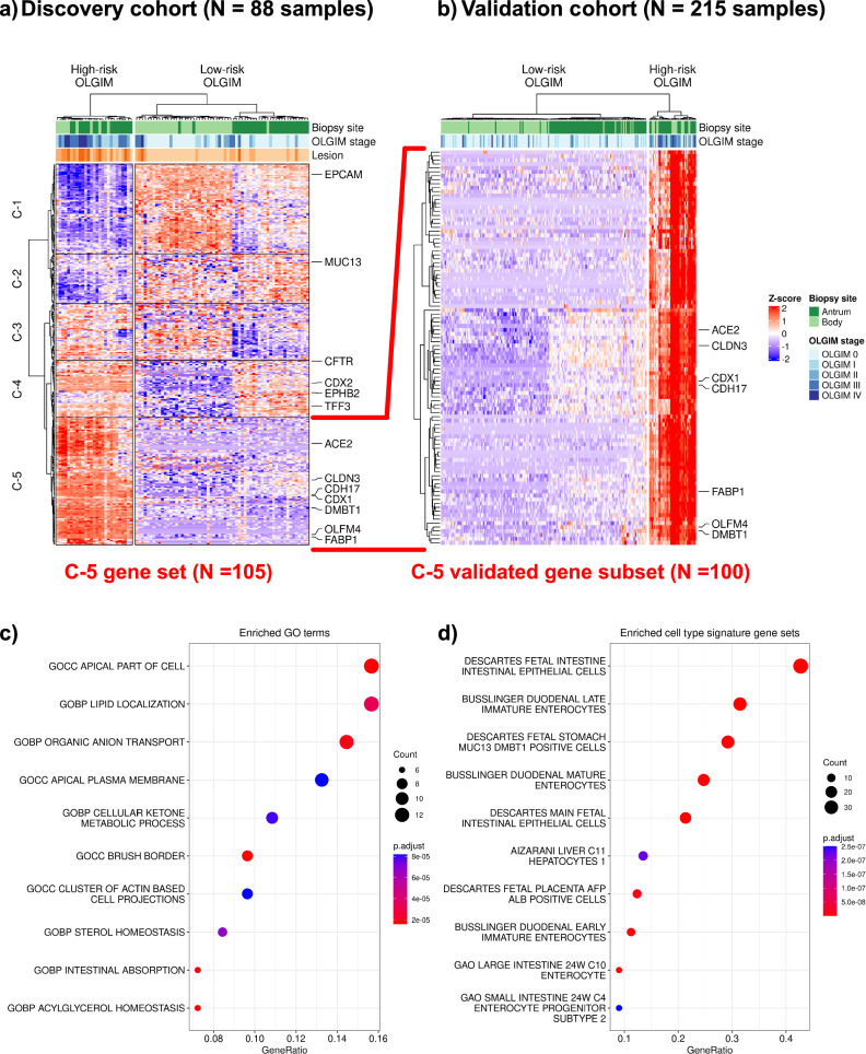
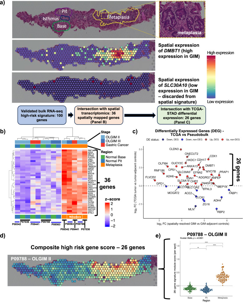
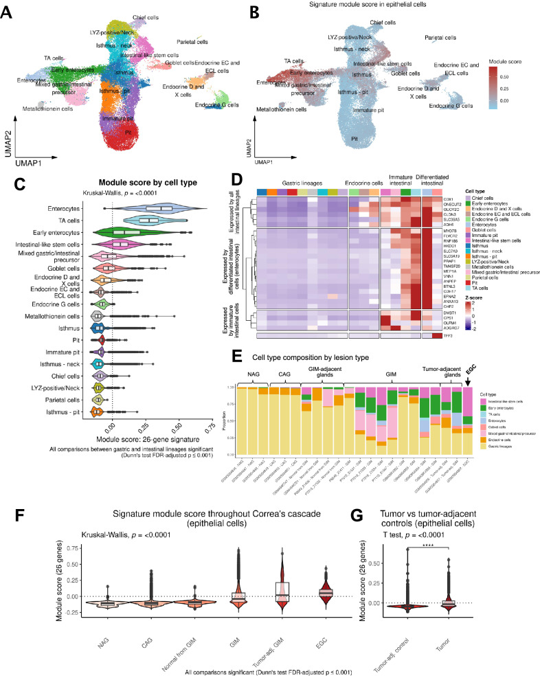
Gastric cancer precursors demonstrate highly-variable rates of progression toward neoplasia. Certain high-risk precursors, such as gastric intestinal metaplasia with advanced histologic features, may be at up to 30-fold increased risk for progression compared to lower-risk intestinal metaplasia. The biological differences between high- and low-risk lesions have been incompletely explored. In this study, we use several clinical cohorts to characterize the microenvironment of advanced gastric cancer precursors relative to low-risk lesions using bulk, spatial, and single-cell gene expression assays. We identified a 26-gene panel which is associated with advanced lesions, localizes to metaplastic glands on histopathology, and is expressed in aberrant mature and immature intestinal cells not normally present in the healthy stomach. This gene expression signature suggests an important role of the immature intestinal lineages in promoting carcinogenesis in the metaplastic microenvironment. These findings may help to inform future biomarker development and strategies of gastric cancer prevention.


Conflict of interest statement

Competing interests: The authors declare no competing interests.

Figures

Fig. 1
Fig. 1. Overview of study design.
Multi-omics flow diagram demonstrating process of discovering and orthogonally validating gene marker panel. At each step, the number of marker genes is shown. This figure was created with BioRender.com.
Fig. 2
Fig. 2. Discovery and validation of the high-risk expression signature.
a Heatmap and hierarchical clustering of differentially expressed and co-expressed genes from the discovery cohort of 88 samples, 22 high-risk (defined as operative link stages III-IV) and 66 low-risk (defined as operative link stages 0-II). Most of the high-risk samples clustered distinctly and separately from the low-risk group, regardless of the anatomic site of the biopsy (top dendrogram). A set of genes were found to be both differentially expressed between high- and low-risk samples. Cluster-5 (C-5) represents 105 genes which were selectively upregulated in their expression only in high-risk samples, regardless of anatomic location. b We found 100 genes from C-5 to be differentially upregulated in the validation cohort (22 high-risk and 193 low-risk samples), confirming a robust signature for high-risk GIM which is agnostic of location. Dot plot depicting over-representation analysis results of these 100 genes: (c) gene ontology terms are enriched with intestinal processes (e.g., brush border, intestinal absorption); (d) cell type signature gene sets are enriched for mature and immature/fetal intestinal cell types.
Fig. 3
Fig. 3. Spatial resolution of the high-risk signature.
a An example of the expression profile of DMBT1 upon a Visium slide annotated by a pathologist for areas of normal glandular architecture (base and pit) and metaplasia. DMBT1 is shown as an example of a spatially resolved gene mapping to pathologist-annotated metaplasia, whereas SLC30A10 is shown as an example of a gene not mapping to metaplasia and, thus, discarded form the spatially-resolved signature. b Heatmap depicting 36 differentially expressed genes from spatial pseudobulk analysis that overlapped with bulk RNA-seq signature from GAPS (FDR-adjusted P value ≤ 0.05; analysis performed using limma-voom). c Scatter plot showing log2 fold-change of 36 upregulated genes from the spatial cohort (X-axis) and log2 fold-change from TCGA. Twenty-six genes overexpressed in both analyses are shown in red. d Spatial mapping of the refined 26-gene signature onto Visium spots. e Comparison of 26-gene signature between metaplastic foci vs normal stomach base or pit (Kruskal-Wallis and Dunn test FDR-adjusted p < 0.001). Note: each Visium spot is 55 µm in diameter, with 100 µm distance between the center of adjacent spots.
Fig. 4
Fig. 4. Single-cell identification of cell types expressing the high-risk signature.
A Uniform Manifold Approximation and Projection (UMAP) plot showing reference-mapped epithelial cells. B UMAP plot showing module score by epithelial cell type. C Comparison of the module score between cell types using all 40 samples from the scRNA-seq cohort. TA, transit amplifying cells. D Heatmap showing the scaled expression of the 26 genes by cell type. E Stacked bar plots depicting the proportion of cell types per sample, ordered by stage of Correa’s cascade. Gastric lineages are aggregated into a single class. EGC Early gastric cancer. F Comparison of module score across Correa’s cascade (p < 0.001 for all comparisons). G Comparison of the module score between GC and tumor-adjacent control tissues (p < 0.0001).
Fig. 5
Fig. 5. Single-molecule fluorescence in situ hybridization (smFISH) of gastric intestinal metaplasia (GIM).
A, B Representative region showing H&E staining of GIM from sample P08563 (operative link III), and superimposed smFISH characterization for six genes: OLFM4, CPS1, HKDC1, DMBT1, ANPEP, and TFF3. C, D Inset magnification for highlighted area from (A) and (B), respectively (90-degree clockwise rotation). E Highlighted regions in (C) and (D), showing H&E and individual channels from areas enriched for intestinal-like stem cells. These cells show elevated expression of OLFM4, DMBT1, CPS1, and HKDC1, together with moderate expression of ANPEP. F Highlighted regions in (C) and (D), showing H&E and individual channels from areas enriched for well-differentiated (mature) cells. These cells show elevated expression of TFF3 and moderate expression of ANPEP. The expression of these genes was mutually exclusive, in space, from the immature cell markers.

Update of

References

    1. Sung, H. et al. Global Cancer Statistics 2020: GLOBOCAN Estimates of Incidence and Mortality Worldwide for 36 Cancers in 185 Countries. CA Cancer J. Clin.71, 209–249 (2021). - PubMed
    1. Allemani, C. et al. Global surveillance of trends in cancer survival 2000-14 (CONCORD-3): Analysis of individual records for 37 513 025 patients diagnosed with one of 18 cancers from 322 population-based registries in 71 countries. Lancet391, 1023–1075 (2018). - PMC - PubMed
    1. Correa, P. Human gastric carcinogenesis: a multistep and multifactorial process–First American Cancer Society Award Lecture on Cancer Epidemiology and Prevention. Cancer Res. 52, 6735–6740 (1992). - PubMed
    1. Sonnenberg, A., Lash, R. H. & Genta, R. M. A national study of Helicobactor pylori infection in gastric biopsy specimens. Gastroenterology139, 1894–1901.e1892 (2010). - PubMed
    1. Sonnenberg, A. & Genta, R. M. Changes in the gastric mucosa with aging. Clin. Gastroenterol. Hepatol.13, 2276–2281 (2015). - PubMed

LinkOut - more resources