. 2025 Feb 25;31(1):78.
doi: 10.1186/s10020-025-01130-8.

A novel mechanism of FTO modulating the progression of endometriosis through mediating the m6A methylation of GEF-H1 in a YTHDF1-dependent manner

Affiliations

A novel mechanism of FTO modulating the progression of endometriosis through mediating the m6A methylation of GEF-H1 in a YTHDF1-dependent manner

Xin-Yu Ding et al. Mol Med. .

Abstract

Background: Endometriosis (EMs) is a condition characterized by the growth of endometrial tissue outside the uterine cavity. Although this condition is benign, it has cancer-like features. N6-methyladenosine (m6A) is a common RNA modification involved in diverse biological processes, but its role in EMs remains unclear.

Methods: A human endometrial stromal cell line (HESCs), primary eutopic endometrial stromal cells (Eu-ESCs), primary ectopic endometrial stromal cells (Ec-ESCs), and clinical samples were used in this study. A colorimetric assay was used to measure methylation levels in clinical and mouse EMs samples. Functional assays (CCK-8, EdU, Transwell, and wound healing) were used to evaluate phenotypic changes. m6A immunoprecipitation sequencing (MeRIP-seq) identified downstream targets. Mechanistic studies were conducted via qRT‒PCR, Western blot, RNA immunoprecipitation (RIP), dual-luciferase reporter, and RNA stability assays.

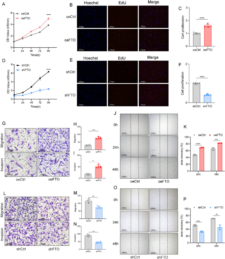
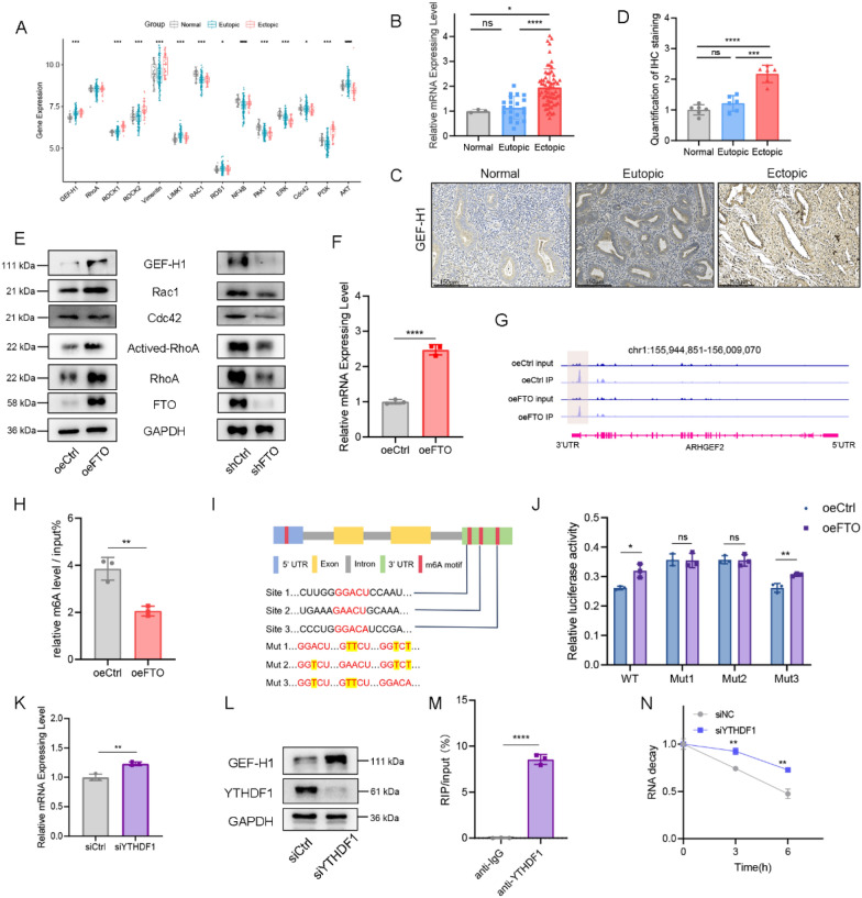
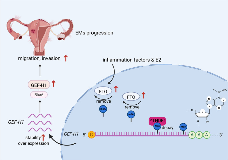
Results: We detected aberrantly low levels of m6A within endometriotic lesions, which was attributed to increased expression of the m6A eraser fat mass and obesity-associated protein (FTO). Notably, estrogen and inflammatory factors, which are recognized as pathogenic agents in EMs amplify FTO expression while suppressing m6A levels. In vitro experiments demonstrated that overexpression of FTO in endometrial stromal cells leads to a reduction in m6A levels and concomitantly promotes their proliferation, migration, and invasion. Furthermore, both genetic deletion of Fto and chemical inhibition of FTO impeded the growth of ectopic endometrial lesions in vivo. By utilizing m6A-seq, we identified GEF-H1 (a Rho guanine nucleotide exchange factor) as a pivotal downstream target of FTO. Specifically, diminished m6A methylation at a certain site within the 3'UTR of GEF-H1 promotes its expression in a YTH N6-methyladenosine RNA-binding protein F1 (YTHDF1)-dependent manner, thereby activating the RhoA pathway. Subsequent experiments revealed that GEF-H1 mediates the effects of FTO in promoting migration and invasion.

Conclusions: This study revealed that FTO decreases the m6A level of GEF-H1, thereby increasing its stability, which in turn activates the GEF-H1-RhoA pathway to promote the migration and invasion of endometrial stromal cells, thereby inducing EMs. Our findings suggest potential therapeutic avenues for targeting FTO to alleviate EMs progression.

Keywords: Endometriosis; Epigenetic modification; FTO; Invasion; Migration; m6A.


Conflict of interest statement

Declarations. Ethics approval and consent to participate: Approval was granted by the ethics committee of the First Affiliated Hospital of Xiamen University. Informed consent was obtained from all individual participants included in the study. Ethics approval number: XMYY-2022KYSB012 Consent for publication: We confirm that all the authors have read and approved the final manuscript for submission. Additionally, all authors consent to the publication of the article. Competing interests: The authors declare no competing interests.

Figures

Fig. 1
Fig. 1
Abnormal m6A modification is observed, and FTO is upregulated in Ems. A The m6A content of total RNA in normal endometrial tissues (Normal; n = 13), eutopic endometrial tissues (Eutopic; n = 13) and paired ectopic endometrial tissues (Ectopic; n = 13) from endometriosis patients. ****P < 0.0001. B The m6A content of total RNA in normal endometria from control mice and ectopic endometria from EMs model mice (n = 7). ***P < 0.001. C Examination of m6A modification-related gene expression in Normal (n = 17), Eutopic (n = 29) and Ectopic (n = 61) samples via qPCR. *P < 0.05, **P < 0.005, ***P < 0.001, ****P < 0.0001. D, E IHC staining of the FTO protein in Normal (n = 6), Eutopic (n = 6), and Ectopic (n = 6) tissues (D), with corresponding quantification of the IHC results (E). Scale bars: 150 μm. ***P < 0.001, ****P < 0.0001. F Scatter plot showing the correlation of the expression of FTO in the ectopic group with the serum CA125 level (n = 29). R = 0.17, P = 0.03. G Scatter plot illustrating the correlation between FTO expression in ectopic lesions and serum AMH levels (n = 21). R = 0.16, P = 0.06. H Overexpression and knockdown efficacy of FTO at the protein level in HESCs. I, J m6A levels of total RNA in HESCs in which FTO was overexpressed or knocked down. ***P < 0.001. KM m6A levels of total RNA in HESCs treated with estrogen, IL-1β or LPS. **P < 0.005, ***P < 0.001. NP qPCR analysis of FTO expression in HESCs treated with various concentrations of estrogen (N), IL-1β (O) or LPS (P). *P < 0.05, **P < 0.005, ***P < 0.001, ****P < 0.0001. Q Immunoblotting assay of FTO expression in HESCs treated with estrogen, IL-1β or LPS
Fig. 2
Fig. 2
FTO regulation of m6A is associated with the proliferation, migration, and invasion of HESCs. AC Cell proliferation was measured by CCK-8 (A) and EdU (B) assays in oeCtrl (negative control) and oeFTO (overexpressing FTO) HESCs. Scale bars: 275 μm. C Quantification of the results of the EdU assay. ****P < 0.0001. DF Cell proliferation was measured by CCK-8 (D) and EdU (E) assays in shCtrl (negative control) and shFTO (knockdown of FTO) HESCs. Scale bars: 275 μm. E Quantification of the results of the EdU assay. ****P < 0.0001. GI Transwell migration and invasion assays of FTO-overexpressing and control HESCs. The results of the quantification of cell migration and invasion are presented in (H, I). ***P < 0.001, ****P < 0.0001. Scale bars: 150 μm. J, K A wound healing assay was performed on HESCs overexpressing FTO. A scratch was created, and the width was measured 24 h and 48 h after the scratch. ***P < 0.001, ****P < 0.0001. Scale bars: 650 μm. LN The invasive and migratory abilities of HESCs transfected with lentiviruses carrying shFTO were explored via a transwell assay. Cells exhibiting migration/invasion are presented in the histogram in (M, N). ***P < 0.001, ****P < 0.0001. Scale bars: 150 μm. O, P Representative images and quantification of the migration of FTO-knockdown HESCs or their corresponding controls. **P < 0.005, ****P < 0.0001. Scale bars: 650 μm
Fig. 3
Fig. 3
Knockout of Fto inhibits ectopic lesion growth in EMs model mice. A Schematic diagram of the EMs mouse model constructed by uterine-specific knockout of Fto. B The expression of FTO in the uteri of control and Ftod/d mice (n = 3). C m6A levels in the uteri of control and Ftod/d mice. ***P < 0.001. D Representative image of ectopic lesions excised from control EMs and Ftod/d EMs mouse models (n = 5). E, F Ectopic lesion weights (E) and ectopic lesion volumes (F) determined after excision from mice in the abovementioned groups. *P < 0.05. (G) m6A levels in HESCs in which FTO was inhibited by Rhein at various concentrations. ***P < 0.001, ****P < 0.0001. H Schematic diagram of the Rhein intervention experiment in the mice with EMs. I Inhibition of FTO effectively suppressed ectopic lesion growth in mice (n = 5). EMs + NS: NS-injected endometriosis group; EMs + Rhein: Rhein-injected endometriosis group. J, K Weights (J) and volumes (K) of ectopic lesions in the EMs and EMs + Rhein groups. ***P < 0.001. L m6A levels of ectopic lesions in the EMs and EMs + Rhein groups. ***P < 0.001
Fig. 4
Fig. 4
MeRIP-Seq identifies GEF-H1 as a downstream target of FTO-mediated m6A modification. A The number of m6A peaks detected in ctrl and oeFTO HESCs. B m6A motif identified in HESCs with or without FTO overexpression. C Distribution of m6A peaks across the length of mRNAs. D, E GO enrichment analysis (D) and KEGG analysis (E) of transcripts with reduced m6A in ctrl versus oeFTO HESCs. (F) Diagram of the Rho pathway with genes affected by m6A marked in yellow. The diagram is based on KEGG annotations
Fig. 5
Fig. 5
FTO-mediated m6A regulates GEF-H1 through YTHDF1. A Box plot of Rho family gene expression in Normal (n = 34), Eutopic (n = 127) and Ectopic (n = 45) samples. *P < 0.05, **P < 0.005, ***P < 0.001, ****P < 0.0001. B GEF-H1 expression levels in Normal (n = 3), Eutopic (n = 21), and Ectopic (n = 67) tissues were analyzed by qPCR. *P < 0.05, ****P < 0.0001. C, D IHC staining (C) and quantitative analysis (D) of GEF-H1 expression in Normal (n = 6), Eutopic (n = 6), and Ectopic (n = 6) tissues. Scale bars: 150 μm. ***P < 0.001, ****P < 0.0001. E Immunoblot analysis of the expression of GEF-H1 and downstream genes in oeFTO and shFTO HESCs. F RT‒qPCR was used to quantify the relative mRNA levels of GEF-H1 transcripts in oeCtrl and oeFTO HESCs. ****P < 0.0001. G The average read density from MeRIP-seq on oeCtrl HESCs and oeFTO HESCs shows the m6A peaks identified in the GEF-H1 transcripts. H MeRIP was used to quantify the relative m6A level of GEF-H1. ***P < 0.001. I Mutations in m6A consensus sequences were generated by replacing adenosine with thymine. J Relative luciferase activity of the wild-type and 3-mutant GEF-H1 3'UTR reporter vectors in FTO-overexpressing HESCs. *P < 0.05, **P < 0.005, ***P < 0.001. K GEF-H1 expression was significantly upregulated at the RNA level in YTHDF1-knockdown HESCs. **P < 0.005. L Immunoblot analysis of the levels of GEH-H1 in HESCs upon transient siRNA knockdown of YTHDF1. ***P < 0.001, ****P < 0.0001. M RIP-qPCR was used to assess the association of the GEF-H1 transcript with the YTHDF1 protein. ****P < 0.0001. N RNA lifetime for GEF-H1 in HESCs transfected with control siRNA or siRNA targeting YTHDF1 was determined by monitoring transcript abundance after transcriptional inhibition. **P < 0.005
Fig. 6
Fig. 6
GEF-H1 mediates the effects of m6A methylation on HESCs migration and invasion. A Effect of GEF-H1 knockdown in HESCs. B Cell proliferation of siCtrl (negative control) and siGEF-H1 (silencing of GEF-H1) HESCs was measured via a CCK-8 assay. C, D EdU assay in HESCs with siCtrl or siGEF-H1 (GEF-H1-knockdown) and quantification of the EdU results. Scale bars: 275 μm. EG Transwell migration and invasion assays of HESCs with siCtrl and siGEF-H1. Scale bars: 275 μm. F, G Cells exhibiting migration/invasion are presented in the histogram. **P < 0.005, ****P < 0.0001. H, I A wound healing assay was performed on HESCs with knockdown of GEF-H1. A scratch was created, and the width was measured 24 h and 48 h after the scratch. ***P < 0.001, ****P < 0.0001. Scale bars: 650 μm. J The efficacy of oeFTO + siGEF-H1 (which inhibits GEF-H1 in HESCs with stable overexpression of FTO) and downstream genes was examined by immunoblotting. KM The invasive and migratory abilities of ctrl, oeFTO and oeFTO + siGEF-H1 HESCs were explored via transwell assays (K) and quantitative analysis (L-M). Scale bars: 275 μm. ***P < 0.001, **** P < 0.0001. (N–O) Representative images and quantification of the migration of GEF-H1-knockdown HESCs or their corresponding controls. Scale bars: 650 μm. *P < 0.05, ***P < 0.001
Fig. 7
Fig. 7
Molecular mechanism by which FTO regulates GEF-H1 degradation through m6A modification, promoting migration and invasion in endometriosis

Similar articles

Cited by

References

    1. Allaire C, Bedaiwy MA, Yong PJ. Diagnosis and management of endometriosis. CMAJ Can Med Assoc J. 2023;195:E363–71. - PMC - PubMed
    1. Becker CM, Gattrell WT, Gude K, Singh SS. Reevaluating response and failure of medical treatment of endometriosis: a systematic review. Fertil Steril. 2017;108:125–36. - PMC - PubMed
    1. Chapron C, Marcellin L, Borghese B, Santulli P. Rethinking mechanisms, diagnosis and management of endometriosis. Nat Rev Endocrinol. 2019;15:666–82. - PubMed
    1. Chen B, Ye F, Yu L, Jia G, Huang X, Zhang X, Peng S, Chen K, Wang M, Gong S, et al. Development of cell-active N6-methyladenosine RNA demethylase FTO inhibitor. J Am Chem Soc. 2012;134:17963–71. - PubMed
    1. Chen L, Gao Y, Xu S, Yuan J, Wang M, Li T, Gong J. N6-methyladenosine reader YTHDF family in biological processes: structures, roles, and mechanisms. Front Immunol. 2023;14:1162607. - PMC - PubMed

MeSH terms

LinkOut - more resources