. 2025 Mar 18;99(3):e0216524.
doi: 10.1128/jvi.02165-24. Epub 2025 Feb 26.

Spike gene variability in porcine epidemic diarrhea virus as a determinant for virulence

Affiliations

Spike gene variability in porcine epidemic diarrhea virus as a determinant for virulence

Wentao Li et al. J Virol. .

Abstract

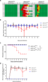
Porcine epidemic diarrhea virus (PEDV) is a pathogenic coronavirus that targets the swine intestinal tract, leading to acute diarrhea and high mortality in neonatal piglets. PEDV is categorized into different genotypes based on genetic variations, especially in the spike (S) gene. The S protein is crucial for viral entry and a major immune target. Significant differences in virulence have been observed among PEDV genotypes, particularly between classical strains and newly emerging strains. In this study, we explored the impact of spike gene variability on PEDV pathogenicity. Using targeted RNA recombination, we generated recombinant PEDV (rPEDV) variants carrying spike genes from contemporary strains (moderately virulent strain UU and highly virulent strain GDU), all within the genetic background of the avirulent DR13 vaccine strain. Pathogenicity was assessed in 3-day-old piglets. The rPEDV carrying the DR13 spike gene was nonpathogenic, with no detectable viral RNA in feces. The rPEDV with the UU spike gene induced mild to severe diarrhea, with moderate viral shedding but no mortality. Conversely, the rPEDV with the GDU spike gene caused severe diarrhea, high viral titers, and high mortality. These findings highlight the critical role of the spike protein in PEDV virulence, informing future development of effective control strategies, including the design of live-attenuated vaccines.IMPORTANCEThis study significantly advances our understanding of how genetic variations in the spike (S) protein of porcine epidemic diarrhea virus (PEDV) influence its ability to cause disease. By engineering viruses with spike genes from different PEDV strains, variations in this protein could be directly linked to differences in disease severity. We found that the spike protein from highly virulent strains caused severe diarrhea and high mortality in piglets, while that from less virulent strains led to milder symptoms. These findings emphasize the central role of the spike protein in determining PEDV virulence, which may enable the design of more effective vaccines to combat PEDV and reduce its impact on the swine industry.

Keywords: PEDV; coronavirus; spike; virulence.


Conflict of interest statement

E.V.D.B., P.J.M.R., and B.-J.B. are listed as inventors of the patents filed on PEDV vaccine strategies. B.N.H., P.V.D.E., and E.V.D.B. are employees of MSD AH. The remaining authors declare no competing interests.

Figures
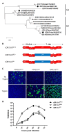
Fig 1
Fig 1
Generation and characterization of recombinant PEDVs. (A) Phylogenetic tree of porcine epidemic diarrhea virus (PEDV) generated using spike gene of representative strains, with strains belonging to the G1 and G2 genogroups indicated. The strains used in this study are highlighted by a black dot (•). (B) Genomes of recombinant DR13 viruses with the spike gene of the DR13 strain (SDR13), the GDU strain (SGDU), or the UU strain (SUU). (C) In vitro characterization of recombinant viruses. Vero cells were infected with the indicated viruses at an MOI of 0.1 in the presence (rDR13-SGDU and rDR13-SUU) or absence or presence (rDR13-SDR13) of 10 µg/mL trypsin. Cells were fixed at 24 hours postinfection and stained with mouse monoclonal antibody 3F12 to the PEDV nucleocapsid protein. Nuclei (blue) were stained with DAPI (blue), and infected cells (green) were visualized by fluorescence microscopy using an anti-PEDV nucleocapsid antibody. (D) Multistep growth kinetics of recombinant PEDV viruses. Vero cells were inoculated with each recombinant PEDV (MOI = 0.01) for 3 hours in the presence (rDR13-SGDU and rDR13-SUU) or absence (rDR13-SDR13) of 10 µg/mL trypsin, after which the inoculum was replaced by fresh culture medium (with or without trypsin). Virus titers were determined at different times postinfection by a quantal assay on Vero cells from which TCID50 values were calculated.
Fig 2
Fig 2
Pathogenicity analysis of recombinant PEDVs in piglets. (A) Diarrhea scores of piglets across different groups over time postinoculation. Scoring criteria: green = normal; yellow = mild diarrhea; red = severe diarrhea; white = severe diarrhea requiring euthanasia or resulting in death. (B) Average rectal temperatures of piglets infected with the rPEDV variants. (C) Kaplan-Meier survival curves displaying the survival rates of piglets infected with the rPEDV variants. The data were analyzed by log-rank test (***, P < 0.001). (D) Fecal PEDV RNA shedding profiles. Data are displayed as mean values ± standard deviations for each group. Values below the detection limit (1.0 log10 GE/mL) were considered negative for viral shedding.
Fig 3
Fig 3
Anti-PEDV-specific IgG and IgA antibody responses following oral inoculation with recombinant PEDVs. The levels of anti-PEDV S1 IgG (A) and IgA (B) antibodies in serum samples collected at different times pre- or postinfection were quantified by ELISA. D-1 represents 1 day before infection; D 12 and D 21 indicate days 12 and 21 postinfection, respectively.

References

    1. Lee C. 2015. Porcine epidemic diarrhea virus: an emerging and re-emerging epizootic swine virus. Virol J 12:193. doi:10.1186/s12985-015-0421-2 - DOI - PMC - PubMed
    1. Mole B. 2013. Deadly pig virus slips through US borders. Nature New Biol 499:388. doi:10.1038/499388a - DOI - PubMed
    1. Sun R-Q, Cai R-J, Chen Y-Q, Liang P-S, Chen D-K, Song C-X. 2012. Outbreak of porcine epidemic diarrhea in suckling piglets, China. Emerg Infect Dis 18:161–163. doi:10.3201/eid1801.111259 - DOI - PMC - PubMed
    1. Chen Q, Gauger PC, Stafne MR, Thomas JT, Madson DM, Huang H, Zheng Y, Li G, Zhang J. 2016. Pathogenesis comparison between the United States porcine epidemic diarrhoea virus prototype and S-INDEL-variant strains in conventional neonatal piglets. J Gen Virol 97:1107–1121. doi:10.1099/jgv.0.000419 - DOI - PubMed
    1. Vlasova AN, Marthaler D, Wang Q, Culhane MR, Rossow KD, Rovira A, Collins J, Saif LJ. 2014. Distinct characteristics and complex evolution of PEDV strains, North America, May 2013-February 2014. Emerg Infect Dis 20:1620–1628. doi:10.3201/eid2010.140491 - DOI - PMC - PubMed

Substances

LinkOut - more resources