. 2025 Jan 21;15(2):158.
doi: 10.3390/biom15020158.

Targeting SMOX Preserves Optic Nerve Myelin, Axonal Integrity, and Visual Function in Multiple Sclerosis

Affiliations

Targeting SMOX Preserves Optic Nerve Myelin, Axonal Integrity, and Visual Function in Multiple Sclerosis

Harry O Henry-Ojo et al. Biomolecules. .

Abstract

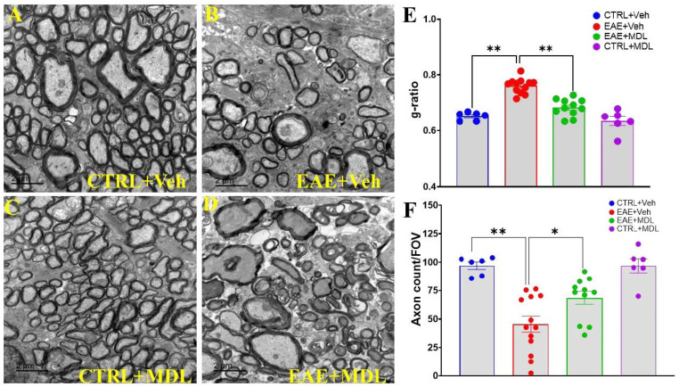
Multiple sclerosis (MS) is a highly disabling chronic neurological condition affecting young adults. Inflammation, demyelination, and axonal damage are key pathological features of MS and its animal model, experimental autoimmune encephalomyelitis (EAE). Our previous work demonstrated that inhibiting spermine oxidase (SMOX) with MDL72527, a selective irreversible pharmacological inhibitor, significantly reduced clinical symptoms, retinal ganglion cell (RGC) loss, and optic nerve inflammation in EAE mice. The present study explored the broader therapeutic potential of SMOX inhibition, focusing on myelin preservation, axonal integrity, and visual function in the EAE model. Electron microscopy of optic nerve cross-sections showed significant preservation of myelin thickness and axonal integrity due to SMOX inhibition. The quantitative assessment showed that g-ratio and axon count metrics were significantly improved in MDL72527-treated EAE mice compared to their vehicle-treated counterparts. Immunofluorescence studies confirmed these findings, showing increased preservation of myelin and axonal proteins in MDL72527-treated EAE mice compared to the vehicle-treated group. Functional assessment studies (Electroretinography) demonstrated significant improvement in RGC function and axonal conduction in EAE mice treated with MDL72527. Furthermore, SMOX inhibition downregulated the expression of galectin3 (Gal3), a mediator of neuroinflammation, indicating Gal3's role in SMOX-mediated neuroprotection. This study provides compelling evidence for the potential of SMOX inhibition as a therapeutic strategy in multiple sclerosis and other demyelinating disorders.

Keywords: MDL72527; multiple sclerosis; myelin; optic nerve; spermine oxidase; vision.


Conflict of interest statement

The authors declare no conflicts of interest.

Figures

Figure 1
Figure 1
MDL72527 treatment attenuates EAE-induced motor deficits. Time course of clinical scores in EAE mice and their respective control groups. Vehicle-treated EAE mice (red line) show progressive worsening of motor function from Day 9 post-induction. However, MDL72527-treated EAE mice (green line) exhibit significantly lower clinical scores. The control groups displayed no motor deficits with scores parallel to the zero line. * p < 0.05 for days 21–27. n = 13–31 mice per group from three different experiments. Data presented as mean ± SEM. Clinical scores are based on a 0–5 scale as described in methods.
Figure 2
Figure 2
Spermine oxidase inhibition preserves optic nerve integrity in EAE mice. (AD) Representative electron microscopy images of optic nerve sections from vehicle control, vehicle-treated EAE, and MDL72527-treated EAE mice. Vehicle-treated EAE mice show axonal loss and demyelination, while MDL72527 treatment markedly reduces these deficits. Scale bar: 2 µm. (E) G-ratios across groups. Vehicle-treated EAE mice show increased g-ratios compared to vehicle control, indicating demyelination. MDL72527 treatment significantly reduces this increase in EAE mice. (F) Axon count per field of view. Vehicle-treated EAE mice exhibit reduced axon counts, while MDL72527 treatment preserves axon integrity. Data presented as mean ± SEM. * p < 0.05, ** p < 0.01. n = 6–13 mice per group.
Figure 3
Figure 3
Axon diameter and g-ratio relationships across experimental groups: Regression plots of inner axon diameter (AD) and outer axon diameter (EH) against g-ratio for WT-CTRL (blue), WT-EAE (red), EAE-MDL (green), and CTRL-MDL (purple). Total g-ratios vs. axon diameter and average g-ratios per mouse across the four mice groups for inner diameter (I,J) and outer diameter (K,L) are shown. Linear regression analysis reveals alterations in both inner and outer axon diameter-g-ratio relationships in EAE and significant improvement with MDL72527 treatment. Inner diameter consistently shows stronger correlation with g-ratio across all groups. Data points represent individual axons; lines indicate linear regression fits. n = 6–13 mice per group.
Figure 4
Figure 4
SMOX inhibition protects against myelin loss in EAE optic nerve. (AD) Representative confocal images of optic nerve sections immunolabeled for myelin basic protein (MBP) in control, vehicle-treated EAE, and MDL72527-treated EAE mice. (A’D’) Magnified images of the boxed regions demonstrating the changes. (E) Quantification of MBP immunofluorescence intensity across groups. (FI) Representative confocal images of optic nerve sections immunolabeled for proteolipid protein (PLP) in control, vehicle-treated EAE, and MDL72527-treated EAE mice. (F’I’) Magnified images of the boxed regions demonstrating the changes. (E) Quantification of MBP immunofluorescence intensity across groups (J) quantification of PLP immunofluorescence intensity across groups. Vehicle-treated EAE mice show substantial loss of both MBP and PLP, while MDL72527 treatment significantly mitigates this loss. Scale bar 50 µm. Data presented as mean ± SEM. ** p < 0.01. n = 5–8 mice per group.
Figure 5
Figure 5
SMOX inhibition rescues EAE-induced axonal injury in the optic nerve. (AD) Representative confocal images of optic nerve sections immune stained for Neurofilament-L (NF-L). (A’D’) Magnified images of the boxed regions demonstrating the changes. (E) Quantification of NF-L immunofluorescence intensity. (FI) Representative confocal images of optic nerve sections immune stained for Neurofilament-M (NF-M). (F’I’) Magnified images of the boxed regions demonstrating the changes. (J) Quantification of NF-M immunofluorescence intensity. (KN) Representative confocal images of optic nerve sections immune stained for beta-III tubulin (TUJ1). (K’N’) Magnified images of the boxed regions demonstrating the changes. (O) Quantification of TUJ1 immunofluorescence intensity. EAE induces downregulation of axonal markers (NF-L, NF-M, TUJ1), which is significantly improved by MDL72527 treatment. Scale bar: 50 µm. Data presented as mean ± SEM. ** p < 0.01. n = 5–8 mice per group.
Figure 6
Figure 6
MDL treatment rescues PERG amplitude in EAE mice and restores RGC electrical signal strength in EAE mice. (A) Composite waveforms for all the mice groups are shown. PERG was recorded in response to reversing gratings (temporal frequency 1 Hz, spatial frequency 0.05 cycles/deg, contrast 1.0). (B) Bar chart displaying P1 and N2 amplitudes across different groups. (C) Bar graph illustrating P1-N1 and P1-N2 amplitudes showing significant differences between WT-EAE mice and MDL-treated groups. Data presented as mean ± SEM; ** p < 0.01. n = 7–8 mice per group. (D) Representative ERG tracings at various intensities. (E) Changes in scotopic b-wave amplitudes were studied at flash intensities ranging from 0.001 to 1.0 cd/s/m2. Data are shown as mean ± SEM. n = 7–8 per group. ** p < 0.01; # p < 0.05.
Figure 7
Figure 7
SMOX inhibition exerts anti-inflammatory effects in EAE disease. (AD) Representative confocal images of optic nerve sections stained with galectin-3 antibody in control, vehicle-treated EAE, and MDL72527-treated EAE mice. (E,F) Gal3 and F4/80 staining on optic nerve sections from Veh EAE and MDL EAE mice showed co-localization, as indicated by the arrows. Scale bar 50 µm. (G) Quantification of galectin immunofluorescence intensity across the groups. Data presented as mean ± SEM. ** p < 0.01. n = 4–6 mice per group.

Similar articles

References

    1. Qian Z., Li Y., Guan Z., Guo P., Zheng K., Du Y., Yin S., Chen B., Wang H., Jiang J., et al. Global, regional, and national burden of multiple sclerosis from 1990 to 2019: Findings of global burden of disease study 2019. Front. Public Health. 2023;11:1073278. doi: 10.3389/fpubh.2023.1073278. - DOI - PMC - PubMed
    1. Lassmann H. Multiple Sclerosis Pathology. Cold Spring Harb. Perspect. Med. 2018;8:a028936. doi: 10.1101/cshperspect.a028936. - DOI - PMC - PubMed
    1. Koch-Henriksen N., Sorensen P.S. The changing demographic pattern of multiple sclerosis epidemiology. Lancet Neurol. 2010;9:520–532. doi: 10.1016/S1474-4422(10)70064-8. - DOI - PubMed
    1. Mike A., Glanz B.I., Hildenbrand P., Meier D., Bolden K., Liguori M., Dell’Oglio E., Healy B.C., Bakshi R., Guttmann C.R. Identification and clinical impact of multiple sclerosis cortical lesions as assessed by routine 3T MR imaging. AJNR Am. J. Neuroradiol. 2011;32:515–521. doi: 10.3174/ajnr.A2340. - DOI - PMC - PubMed
    1. Gelfand J.M. Handbook of Clinical Neurology. Volume 122. Elsevier; Amsterdam, The Netherlands: 2014. Multiple sclerosis: Diagnosis, differential diagnosis, and clinical presentation; pp. 269–290. - DOI - PubMed

MeSH terms

LinkOut - more resources