. 2025 Feb 15;15(2):293.
doi: 10.3390/biom15020293.

Lipid-siRNA Conjugates Targeting High PD-L1 Expression as Potential Novel Immune Checkpoint Inhibitors

Affiliations

Lipid-siRNA Conjugates Targeting High PD-L1 Expression as Potential Novel Immune Checkpoint Inhibitors

Rina Tansou et al. Biomolecules. .

Abstract

Programmed death 1 ligand (PD-L1), an important immune checkpoint molecule, is mainly expressed on cancer cells and has been shown to exert an immunosuppressive effect on T-cell function by binding to programmed cell death 1 (PD-1) expressed on T-cells. Recently, immune checkpoint inhibitors using antibody drugs such as nivolumab and atezolizumab have attracted attention. However, clinical challenges, including limitations to the scope of their application, are yet to be addressed. In this study, we developed a novel immune checkpoint inhibitor that targets PD-L1 using lipid-siRNA conjugates (lipid-siPDL1s). The inhibitory effect of lipid-siPDL1s on PD-L1 expression was evaluated and found to strongly suppress mRNA expression. Notably, lipid-siPDL1s exerted a significantly stronger effect than unmodified siPDL1. Interestingly, lipid-siPDL1s strongly inhibited PD-L1 expression despite cancer cell stimulation by interferon-gamma, which induced the overexpression of PD-L1 genes. These results strongly suggest that lipid-siPDL1s could be used as novel immune checkpoint inhibitors.

Keywords: immune checkpoint inhibitors; interferon gamma (IFNγ) stimulation; nucleic acid drugs; programmed death 1 ligand (PD-L1); siRNA conjugates.


Conflict of interest statement

The authors declare no conflicts of interest.

Figures

Figure 1
Figure 1
Synthesis (A), reverse-phase high-performance liquid chromatography (RP-HPLC) profiles (B), sequences and structures (C), and polyacrylamide gel electrophoresis (PAGE) analysis of lipid-siPDL1s targeting PD-L1 mRNA (D). Lipid-ssPDL1s were synthesized using a simple conjugation method [43]. Fatty acids were conjugated to the 5′-end of the sense strand via an amino linker. RP-HPLC was performed using an octadecylsilyl column (4.6 × 75 mm I.D., 5 μm) and a linear gradient of acetonitrile at concentrations varying from 7 to 70% over 40 min in 100 mM triethylamine acetate (pH 7.0). The elution time depends on the nature of conjugated lipids. The purified lipid-ssPDL1 is double-stranded with the antisense strand, and PAGE analysis confirmed sufficient purity of the resulting lipid-siPDL1s. Original western blot images can be found at Supplementary Materials.
Figure 2
Figure 2
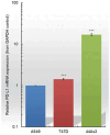
Relative PD-L1 mRNA expression in A549, T47D, and 44As3 cells. PD-L1 mRNA was evaluated in A549 cells and compared with that in T47D and 44As3 cell lines using RT-qPCR. Cancer cell lines exhibit differential PD-L1 expression. Comparing the three cancer cell lines, PD-L1 expression is the highest in 44As3 cells. Data are presented as the mean ± SD of three independent experiments (*** p < 0.001 vs. A549; t-test). PD-L1, Programmed death 1 ligand; RT-qPCR, reverse transcription-quantitative PCR.
Figure 3
Figure 3
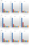
RNAi efficacy of siPDL1 and lipid-siPDL1s against A549 (A), T47D (B), and 44As3 (C) cells. The RNAi effects of siPDL1 and lipid-siPDL1s vary across the different cell types. siPDL1 and lipid-siPDL1s suppress PD-L1 expression by 60–80% in A549, 60–90% in T47D, and 50–80% in 44As3 cells compared to siCtrl-treated cells. For all cell lines, lipid-siPDL1s exhibit stronger RNAi effects than siPDL1. Data are presented as the mean ± SD of 3–5 independent experiments (** p < 0.01, *** p < 0.001 vs. siPDL1; t-test). PD-L1, Programmed death 1 ligand; RNAi, RNA interference.
Figure 4
Figure 4
Alterations in PD-L1 expression in A549, T47D, and 44As3 cell lines following IFNγ stimulation. A549, T47D, and 44As3 cells exhibit a 5- to 20-fold increase in PD-L1 expression upon IFNγ stimulation (A). Among the three cell types, 44As3 cells exhibit the highest PD-L1 mRNA expression (B), even upon IFNγ stimulation. Western blot analysis (C) demonstrates increased PD-L1 protein expression in each cancer cell line following IFNγ stimulation. Data are presented as the mean ± SD of three independent experiments (*** p < 0.001 vs. A549; t-test). IFNγ, interferon-gamma; PD-L1, Programmed death 1 ligand. Original western blot images can be found at Supplementary Materials.
Figure 5
Figure 5
RNAi efficacies of siPDL1 and lipid-siPDL1s in A549 (A), T47D (B), and 44As3 (C) cell lines upon IFNγ-induced PD-L1 upregulation. Both siPDL1 and lipid-siPDL1s exhibit significant RNAi effects on cancer cells with high PD-L1 expression levels at 24–72 h post-treatment. siPDL1 and lipid-siPDL1s suppress PD-L1 expression by 60–90% in A549, 40–80% in T47D, and 60–80% in 44As3 cells compared with that in siCtrl-treated cells. Lipid-siPDL1s exert stronger RNAi effects than siPDL1 under all experimental conditions, with variations depending on IFNγ sensitivity. Data are presented as the mean ± SD of 3–5 independent experiments (ns = not significant, * p < 0.05, ** p < 0.01, *** p < 0.001 vs. siPDL1; t-test). IFNγ, interferon-gamma; PD-L1, Programmed death 1 ligand.
Figure 6
Figure 6
Confocal microscopy analysis of the effects of lipid-siPDL1s (50 nM) knockdown on IFNγ-stimulated PD-L1 protein expression in A549 (A), T47D (B), and 44As3 (C) cells after 48 and 96 h. Although cancer cells were stimulated with IFNγ (100 ng/mL) and exhibited high PD-L1 expression, siPDL1 and lipid-siPDL1s suppress the surface PD-L1 protein expression in these cells. In particular, lipid-siPDL1s strongly suppress cell surface PD-L1 protein, and the effect is long-lasting. Blue fluorescence (Hoechst33342) indicates nuclei; green fluorescence (Alexa-488 labeled anti-PD-L1 antibody) indicates PD-L1 on cancer cells; red fluorescence (MitoTracker Red) indicates mitochondria, and the merged images show the overlay of blue, green, and red fluorescence. IFNγ, interferon-gamma; PD-L1, Programmed death 1 ligand.
Figure 7
Figure 7
Western blot analysis of the inhibitory effect of lipid-siPDL1s on PD-L1 protein expression in A549 (A), T47D (B), and 44As3 (C) cells overexpressing PD-L1. Cells treated with siCtrl show strong expression of PD-L1 protein, accompanied by the detection of a strong signal; however, cells treated with lipid-siPDLs exhibit weaker expression of PD-L1 protein, and the effects of mRNA knockdown correlating with the suppression of PD-L1 protein expression. Original western blot images can be found at Supplementary Materials.

Similar articles

Cited by

References

    1. Borkner L., Kaiser A., van de Kasteele W., Andreesen R., Mackensen A., Haanen J.B., Schumacher T.N., Blank C. RNA interference targeting programmed death receptor-1 improves immune functions of tumor-specific T cells. Cancer Immunol. Immunother. 2010;59:1173–1183. doi: 10.1007/s00262-010-0842-0. - DOI - PMC - PubMed
    1. Lian S., Xie R., Ye Y., Lu Y., Cheng Y., Xie X., Li S., Jia L. Dual blockage of both PD-L1 and CD47 enhances immunotherapy against circulating tumor cells. Sci. Rep. 2019;9:4532. doi: 10.1038/s41598-019-40241-1. - DOI - PMC - PubMed
    1. Sanmamed M.F., Chen L. A Paradigm Shift in Cancer Immunotherapy: From Enhancement to Normalization. Cell. 2018;175:313–326. doi: 10.1016/j.cell.2018.09.035. - DOI - PMC - PubMed
    1. Bolandi N., Derakhshani A., Hemmat N., Baghbanzadeh A., Asadzadeh Z., Afrashteh Nour M., Brunetti O., Bernardini R., Silvestris N., Baradaran B. The Positive and Negative Immunoregulatory Role of B7 Family: Promising Novel Targets in Gastric Cancer Treatment. Int. J. Mol. Sci. 2021;22:10719. doi: 10.3390/ijms221910719. - DOI - PMC - PubMed
    1. Blank C., Brown I., Peterson A.C., Spiotto M., Iwai Y., Honjo T., Gajewski T.F. PD-L1/B7H-1 inhibits the effector phase of tumor rejection by T cell receptor (TCR) transgenic CD8+ T cells. Cancer Res. 2004;64:1140–1145. doi: 10.1158/0008-5472.CAN-03-3259. - DOI - PubMed

MeSH terms