. 2025 Feb 3;14(2):155.
doi: 10.3390/biology14020155.

Pigment Epithelium-Derived Factor Inhibits Cell Motility and p-ERK1/2 Signaling in Intrahepatic Cholangiocarcinoma Cell Lines

Affiliations

Pigment Epithelium-Derived Factor Inhibits Cell Motility and p-ERK1/2 Signaling in Intrahepatic Cholangiocarcinoma Cell Lines

Veronica Porreca et al. Biology (Basel). .

Abstract

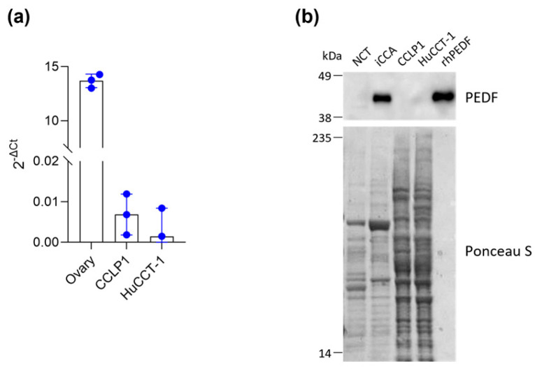
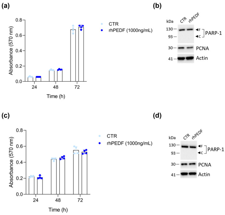
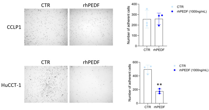
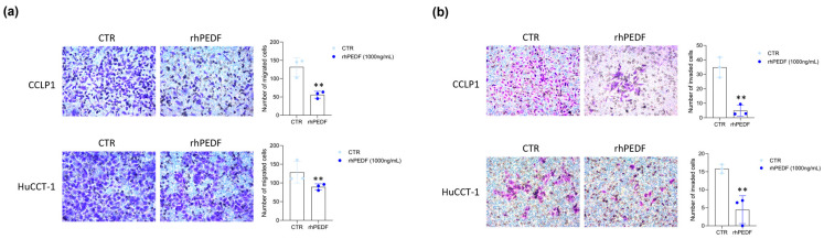
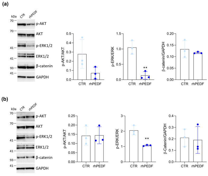
Pigment epithelium-derived factor (PEDF) is a multifunctional soluble glycoprotein, primarily known for its potent anti-angiogenic properties. In recent years, its ability to counteract cell proliferation and motility has generated interest in PEDF as a potential tumor suppressor. In the intrahepatic Cholangiocarcinoma (iCCA), PEDF, Thrombospondin 1 (THBS1), and Thrombospondin 2 (THBS2) are expressed and released into the tumor microenvironment (TME), where they promote lymphangiogenesis at the expense of the neoangiogenic program, aiding the dissemination of cancer cells via lymphatic vessels. Recently, we demonstrated that THBS1 and THBS2 directly affect iCCA cells, exacerbating their malignant behavior, while the direct role of PEDF remains to be elucidated. In this study, through a cell-based assay and molecular analysis, we investigate the direct function of PEDF on two well-established iCCA cell lines. Our results show that PEDF affects cancer cell motility in a paracrine manner, reducing their migratory and invasive capabilities. Notably, our data suggest that the PEDF-induced inhibition of motility in iCCA cells occurs through the MAPK/ERK signaling pathway, as indicated by the reduced phosphorylation of ERK1/2. Overall, this study provides the first evidence of PEDF acting as a tumor suppressor in iCCA.

Keywords: PEDF; cell migration; extracellular matrix; intrahepatic cholangiocarcinoma; tumor microenvironment.


Conflict of interest statement

The authors declare no conflicts of interest.

Figures

Figure 1
Figure 1
CCLP1 and HuCCT-1 cell lines do not express PEDF. (a) PEDF mRNA expression in iCCA cell lines, (CCLP1 and HuCCT-1) was analyzed using Real-Time PCR. Commercial human ovary RNA was used as a positive control. The bar graph illustrates the mean 2−ΔCt of PEDF mRNA normalized with respect to the expression of GAPDH. (b) Western blot analysis for PEDF in total tissue extracts from non-cancerous (NCT) and cancerous tissues (iCCA) of a representative iCCA patient as well as total cell extracts from CCLP1 and HuCCT-1 cell lines. For each gel lane, 10 μg of protein were loaded. A total of 10 ng of rhPEDF was used as a positive control. Ponceau S staining of the nitrocellulose membrane is shown to confirm equal protein loading.
Figure 2
Figure 2
In vitro cell growth is not influenced by rhPEDF treatment. The time course of cell growth for CCLP1 (a) and HuCCT-1 (c) cell lines was assessed by MTT cell viability assay at the indicated time points in the presence of 1000 ng/mL of rhPEDF; CTR represents untreated cells. Western blot analysis for PCNA and PARP-1 on whole cell extracts of CCLP1 (b) and HuCCT-1 (d) cells, cultured in the presence of 1000 ng/mL of rhPEDF for 24 h, was performed. Arrows indicate the expected molecular weights for the PARP-1 full-length “F” and cleaved “C” forms; actin was used as a loading control.
Figure 3
Figure 3
rhPEDF inhibits the cellular adhesive properties of the epithelial HuCCT-1 cell line. Adhesion of CCLP1 and HuCCT-1 cells in response to treatment with (rhPEDF) or without (CTR) 1000 ng/mL of rhPEDF for 45 min. The left panels show representative images (4× magnification) of CCLP1 and HuCCT-1 adherent cells. The bar graphs represent the mean value ± SD of cell counts per four optical fields of three independent experiments ** p < 0.01 (t-test).
Figure 4
Figure 4
rhPEDF treatment restrains cell migration and invasion in iCCA cell lines. Transwell migration (a) and invasion (b) assay of CCLP1 and HuCCT-1 cells in response to treatment with (rhPEDF) or without (CTR) 1000 ng/mL of rhPEDF for 48 h. The left panels show representative images (20× magnification) of each experimental group. The bar graphs represent the mean value ± SD from independent experiments performed in triplicate; ** p < 0.01 (t-test).
Figure 5
Figure 5
rhPEDF downregulates ERK1/2 phosphorylation. Western blot analysis for the indicated proteins on whole cell extracts of CCLP1 (a) and HuCCT-1 (b) cells, cultured in response to treatment with (rhPEDF) or without (CTR) 1000 ng/mL of rhPEDF for 24 h; GAPDH was used as a loading control. Quantification from three independent immunoblots by densitometry analysis is shown (for details see Section 2); ** p < 0.01 (t-test).

Similar articles

Cited by

References

    1. Steele F.R., Chader G.J., Johnson L.V., Tombran-Tink J. Pigment epithelium-derived factor: Neurotrophic activity and identification as a member of the serine protease inhibitor gene family. Proc. Natl. Acad. Sci. USA. 1993;90:1526–1530. doi: 10.1073/pnas.90.4.1526. - DOI - PMC - PubMed
    1. Tombran-Tink J., Barnstable C.J. PEDF: A multifaceted neurotrophic factor. Nat. Rev. Neurosci. 2003;4:628–636. doi: 10.1038/nrn1176. - DOI - PubMed
    1. Tombran-Tink J., Mazuruk K., Rodriguez I.R., Chung D., Linker T., Englander E., Chader G.J. Organization, evolutionary conservation, expression and unusual Alu density of the human gene for pigment epithelium-derived factor, a unique neurotrophic serpin. Mol. Vis. 1996;2:11. - PubMed
    1. Akiba J., Yoshida T., Sadashima E., Murata K., Matsui T., Yamagishi S.I., Kusano H., Mihara Y., Mizuochi S., Kinjou Y., et al. The Expression of PEDF and its Putative Receptors in Hepatocellular Carcinoma and Background Liver Tissue. Anticancer. Res. 2021;41:1203–1212. doi: 10.21873/anticanres.14877. - DOI - PubMed
    1. Petersen S.V., Valnickova Z., Enghild J.J. Pigment-epithelium-derived factor (PEDF) occurs at a physiologically relevant concentration in human blood: Purification and characterization. Biochem. J. 2003;374:199–206. doi: 10.1042/bj20030313. - DOI - PMC - PubMed

LinkOut - more resources